Microscope Magnification Calculator
Magnification is image size ÷ actual size in the same unit. A microscope’s total magnification is eyepiece × objective. Work out both, plus field of view.
Calculator
Magnification, image size and actual size
M = I ÷ A, with both sizes in the same unit. Choose the one to solve for and fill in the other two.
Measured on the drawing, photograph or micrograph.
The real size of the specimen.
Of the image you measured. For a drawing that is not the lens setting.
Working, with your numbers
- M = I / A
- = 45 mm / 90 µm
- = 45,000 µm / 90 µm
- = ×500
Both sizes are put into one unit first, because millimetres divided by micrometres as bare numbers put the answer out by a factor of 1000.
- 1 mm on the image 1 mm ÷ M: how much of the specimen one millimetre of this image covers, for reading sizes straight off it.
- 2 µm
- Scale bar A round length no longer than the specimen, and how long to draw it on the image at this magnification. A scale bar stays right when the image is resized; a stated magnification does not.
- 50 µm drawn 25 mm long
Total magnification, field of view and cell size
Read the lens settings off the microscope. The field of view comes from the eyepiece’s field number, or from a field you measured yourself at another magnification.
Engraved on the eyepiece, often with the field number: 10×/18.
Engraved on the objective barrel, such as 4, 10, 40 or 100.
1 unless the microscope adds a tube factor or a magnification changer, such as 1.25 or 1.5.
The number after the slash on the eyepiece: 18 on a 10×/18.
How many cells, end to end, span the diameter. Fractions are fine.
Microscope working
- Total magnification = 10 × 40 = ×400
- Field of view = 18 mm / 40 = 0.45 mm = 450 µm
- Cell size = 450 µm / 5 = 90 µm
The field of view depends on the objective, any extra factor and the field number, not on the eyepiece magnification.
- Total magnification Eyepiece × objective × any extra factor. It describes the lenses, not a drawing made through them.
- ×400
- Field of view Field number ÷ (objective × extra factor). The eyepiece magnification does not enter it.
- 0.45 mm = 450 µm
- Field area π × (diameter ÷ 2)². Divide a count of cells or stomata in one field by this for a number per mm².
- 0.159 mm²
- Estimated cell size Field of view ÷ cells across. An average over the cells you counted, and only as good as the count.
- 90 µm
Citing this tool
Last updated . Add the date you accessed it as well, which a citation of a page that can change asks for. If a specific result matters, cite the permalink from the tool’s share row instead of this page: it reproduces the exact parameters.
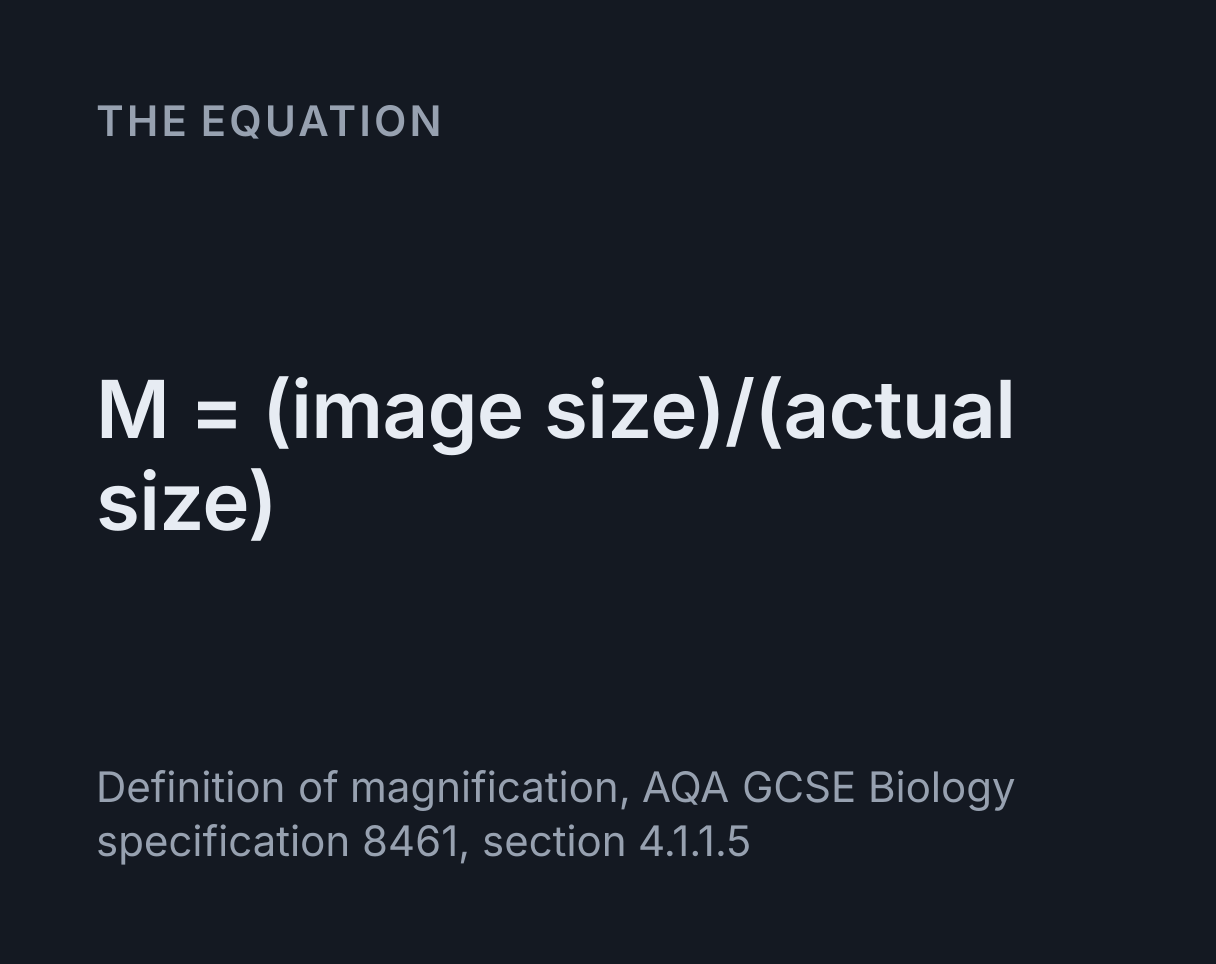
The equation
Definition of magnification, AQA GCSE Biology specification 8461, section 4.1.1.5
How to calculate magnification
Magnification is the size of the image divided by the actual size of the specimen, with both
lengths in the same unit: M = I ÷ A, or magnification = image size ÷ actual
size. Rearranged, the actual size is A = I ÷ M and the image size is
I = M × A, so any two of the three give the third. The AQA GCSE and A-level
biology specifications write it the same way. A microscope’s total magnification is a
separate number, eyepiece × objective, which the second panel works out.
Magnification has no unit, because it is one length divided by another, and it is written with a multiplication sign, as ×500. It only comes out right when the two lengths share a unit. Images are measured with a ruler in millimetres and cells are quoted in micrometres, and 1 mm = 1000 µm, so a size in millimetres is multiplied by 1000 before it is divided by one in micrometres. Electron micrographs take the same step one prefix further down, 1 µm = 1000 nm, and the SI prefixes table has the rest.
Using the calculator
The first panel solves M = I ÷ A for whichever term you mark, with a unit menu on
each size, and prints the working with the conversion written out. Under it, “1 mm on the
image” is the length of specimen one millimetre of the image covers, and “Scale bar” gives a
round length no longer than the specimen and how long to draw it at that magnification.
The second panel takes the lens settings: the total magnification, the diameter and area of the field of view, and a cell size estimated from how many cells span the field. The two panels are kept apart on purpose, because the magnification of a drawing or a photograph is not the magnification of the lenses it was made through.
Worked example: the magnification of a drawing
A cell is drawn 45 mm wide, and its real width is 90 µm. What is the magnification of the drawing?
- Put the image size in micrometres:
45 mm × 1000 = 45,000 µm M = I ÷ A = 45,000 µm ÷ 90 µmM = ×500
Going the other way, an electron micrograph at ×12,000 shows a bacterium
36 mm long. Its actual length is 36 mm ÷ 12,000 = 0.003 mm,
which is 3 µm. Above ×1, an actual size larger than the image means the
division was done the wrong way up.
Total magnification of a microscope
A compound light microscope magnifies in two stages. The objective forms an enlarged image
inside the tube and the eyepiece enlarges that image again, so the total is the product:
total magnification = eyepiece × objective. A ×10 eyepiece on a ×40 objective
gives ×400, and on a ×100 oil immersion objective ×1000. Some microscopes add a tube factor or
a magnification changer, which multiplies in the same way: Evident’s microscopy primer works
through a ×5 objective with ×15 eyepieces and a 1.25× tube factor, for ×93.75. Each stage is a
converging lens forming an image, which the thin lens
calculator solves and the ray diagram simulator
draws.
That figure describes the lenses, not anything drawn through them. The 90 µm cell above, seen at ×400 and drawn 45 mm wide, makes a drawing at ×500, because a drawing’s magnification is set by how large it is drawn. Label a drawing with its own magnification or with a scale bar.
Field of view and cell size
The field of view is the diameter of the circle of specimen you can see. It is set by the
eyepiece’s field number, the diameter in millimetres of the fixed diaphragm
inside it, which is engraved after its magnification: a 10×/18 eyepiece has a field number of
18. Dividing by the objective magnification, and by any tube factor, gives the field at the
specimen: field of view = FN ÷ (objective × factor). The eyepiece magnification
is not in it.
With a 10×/18 eyepiece the field is 18 ÷ 4 = 4.5 mm across with a ×4 objective,
1.8 mm with ×10, 0.45 mm with ×40 and 0.18 mm with ×100. To estimate the size of a cell, count
how many span the diameter and divide: five cells across the 0.45 mm field, which is 450 µm,
are about 90 µm each. That is an estimate rather than a measurement, an
average over the cells counted.
If the eyepiece shows no field number, measure the field at low power with a clear ruler and
scale it. With the same eyepiece the field’s diameter is inversely proportional to the total
magnification, so 4.5 mm measured at ×40 becomes 4.5 × 40 ÷ 400 = 0.45 mm at
×400. Choose A field I measured above and the calculator does that step.
Common mistakes
- Dividing sizes in different units. 45 mm ÷ 90 µm as bare numbers gives 0.5, a thousand times too small. Convert first: 1 mm = 1000 µm and 1 µm = 1000 nm.
- Using the lens setting as a drawing’s magnification. A cell seen at ×400 can be drawn at ×500 or at ×50, depending on how large it is drawn. The drawing’s magnification is its size over the cell’s.
- Multiplying by the magnification to get the actual size. The actual size is the image size divided by M, so above ×1 it always comes out smaller than the image.
- Adding the lens magnifications. A ×10 eyepiece and a ×40 objective make ×400, not ×50.
- Measuring the high power field with a ruler. At ×400 the field is about 0.45 mm across, narrower than one millimetre division, so measure at low power and scale it.
What this calculator leaves out
Resolution. Magnification says how large the image is, not how much detail it holds. An ordinary light microscope cannot resolve detail finer than about 0.2 µm, which is Abbe’s limit, wavelength ÷ (2 × numerical aperture), for green light of 550 nm through an oil immersion objective of numerical aperture 1.4. Past about 1000 times the numerical aperture, roughly ×1400 for that objective, the image only gets bigger. The calculator warns when the lens settings pass that, and when an actual size falls below 0.2 µm.
Graticule calibration. At A-level, cells are measured with an eyepiece
graticule calibrated against a stage micrometer: if 40 graticule divisions line up with
100 µm of the stage micrometer, each division is 100 ÷ 40 = 2.5 µm with that
objective. The calculator does not do that step, but a size worked out that way goes straight
into the actual size field.
Photographs and screens. A magnification printed beside an image is only right at the size it was printed. Once a micrograph is zoomed on a screen or resized, measure against its scale bar instead, which changes size with it.
Converting units first? Use the length conversion table.
Common questions
How do you calculate magnification in biology?
Divide the image size by the actual size, after putting both in the same unit: magnification = image size ÷ actual size. A cell drawn 45 mm wide that is really 90 µm across is at 45,000 µm ÷ 90 µm = ×500. Rearranged, actual size = image size ÷ magnification, and image size = magnification × actual size.
How do you calculate the actual size of a specimen?
Divide the image size by the magnification: actual size = image size ÷ magnification. A bacterium measuring 36 mm on an electron micrograph at ×12,000 is really 36 mm ÷ 12,000 = 0.003 mm long, which is 3 µm. Measure the image in millimetres and convert the answer to micrometres, the unit cells are usually quoted in.
How do you work out the total magnification of a microscope?
Multiply the eyepiece magnification by the objective magnification. A ×10 eyepiece with a ×40 objective gives ×400, and with a ×100 oil immersion objective ×1000. If the microscope has a tube factor or a magnification changer, multiply that in as well: ×10, ×40 and a 1.25× factor give ×500.
How do you calculate the field of view of a microscope?
Divide the eyepiece’s field number by the objective magnification. A 10×/18 eyepiece has a field number of 18 mm, so with a ×40 objective the field of view is 18 ÷ 40 = 0.45 mm, or 450 µm, across. If you measured the field at low power instead, scale it by the ratio of the two magnifications, low power over high: 4.5 mm at ×40 is 0.45 mm at ×400 with the same eyepiece.
How do you estimate the size of a cell from the field of view?
Divide the diameter of the field of view by the number of cells that span it. Five cells across a 450 µm field are about 90 µm each. It is an estimate rather than a measurement, because it averages the cells you counted and assumes they lie end to end across the middle of the field.
Does a higher magnification always show more detail?
No. Past about 1000 times the objective’s numerical aperture the image gets bigger without showing more detail, which is called empty magnification. Most oil immersion objectives stop at a numerical aperture of 1.4, so about ×1400 is the useful ceiling, and no magnification lets an ordinary light microscope resolve detail finer than about 0.2 µm.