Cell Structure Explorer
Labelled animal and plant cell diagrams: tap any organelle for its structure and function, compare the two cells, and see how big each part really is.
Visualiser
Tap a part of the drawing or choose it from the key. Small parts are drawn larger than life, so the size ladder below gives their real sizes.
Nucleus selected.
Animal cell
Nucleus
Found in: Animal and plant cells.
Structure
Usually the most prominent organelle. It is bounded by the nuclear envelope, two membranes pierced by nuclear pores and continuous with the ER, and filled with nucleoplasm holding chromatin: DNA wound with proteins such as histones.
Function
Holds the cell’s DNA and directs the making of ribosomes and proteins. The pores control what passes between the nucleus and the cytoplasm, including the RNA that carries each gene’s instructions out.
Size
Typically 2 to 10 µm across. Source: Milo and Phillips, Cell Biology by the Numbers (2015).
Size ladder
Typical sizes on a scale where each step is ten times the last. Parts to the left of the dashed line, at 200 nm, are too small for a light microscope to show; an electron microscope is needed.
- Animal or plant cell 10 to 100 µm
- Nucleus 2 to 10 µm
- Red blood cell 7 to 8 µm
- Chloroplast 4 to 6 µm
- Bacterium 100 nm to 5 µm
- Mitochondrion 1 to 2 µm
- ER tubule 30 to 100 nm
- Ribosome 20 to 30 nm
- Cytoskeleton fibre 7 to 25 nm
- Membrane thickness 4 to 10 nm
Sources: Milo and Phillips, Cell Biology by the Numbers (2015), for the organelles and the red blood cell; OpenStax Biology 2e (Clark, Douglas and Choi, 2018), chapter 4, for cells, bacteria, the cytoskeleton and the microscope limit.
Animal and plant cells compared
| Part | Animal cell | Plant cell |
|---|---|---|
| Cell membrane | Yes | Yes |
| Cytoplasm | Yes | Yes |
| Nucleus | Yes | Yes |
| Nucleolus | Yes | Yes |
| Ribosomes | Yes | Yes |
| Rough endoplasmic reticulum | Yes | Yes |
| Smooth endoplasmic reticulum | Yes | Yes |
| Golgi apparatus | Yes | Yes |
| Vesicles | Yes | Yes |
| Mitochondria | Yes | Yes |
| Lysosomes | Yes | Most do not |
| Peroxisomes | Yes | Yes |
| Centrosome | Yes | Most do not |
| Cytoskeleton | Yes | Yes |
| Cell wall | No | Yes |
| Chloroplasts | No | Yes |
| Central vacuole | No | Yes |
| Plasmodesmata | No | Yes |
Citing this tool
Last updated . Add the date you accessed it as well, which a citation of a page that can change asks for. If a specific result matters, cite the permalink from the tool’s share row instead of this page: it reproduces the exact parameters.
The equation
Surface area and volume of a sphere, as in OpenStax Biology 2e (2018), section 4.2
The parts of a cell and what they do
A cell is a set of organelles, each with its own job: the nucleus holds the DNA, ribosomes build proteins, the endoplasmic reticulum and the Golgi apparatus finish and ship them, and mitochondria release the energy in food as ATP. Animal and plant cells share all of these. A plant cell adds a cellulose cell wall, chloroplasts and a large central vacuole, and most plant cells lack the centrosome and the lysosomes an animal cell has.
The explorer draws both cells, 14 parts in the animal cell and 16 in the plant cell, as original labelled diagrams arranged the way a textbook arranges them. Choose a part to see what it is made of, what it does and how big it really is.
Using the explorer
- Switch cells with the buttons above the drawing. The key under it numbers the parts of whichever cell is showing.
- Choose a part by tapping it in the drawing or pressing its button in the key. The arrow keys step through the key in order, so the whole cell can be toured from the keyboard.
- Read the card beside the drawing: where the part is found, its structure, its function, and its size with the source of the figure.
- Choose a part the cell does not have, such as chloroplasts in the animal cell, and the card says so and what the cell does instead. That is the quickest way to learn the differences, and a button takes you to the cell that has it.
Animal cells and plant cells compared
Parts found in both kinds of cell: cell membrane, cytoplasm, nucleus, nucleolus, ribosomes, rough endoplasmic reticulum, smooth endoplasmic reticulum, Golgi apparatus, vesicles, mitochondria, peroxisomes and cytoskeleton.
Parts found only in the animal cell: lysosomes and centrosome. OpenStax Biology puts it as “animal cells each have a centrosome and lysosomes; whereas, most plant cells do not”: a plant cell’s vacuole does the digesting a lysosome does, and plant cells organise their microtubules without centrioles.
Parts found only in the plant cell: cell wall, chloroplasts, central vacuole and plasmodesmata. The wall is why plant cells keep a fixed, often box-like shape while animal cells are rounder and more varied, and a full vacuole pressing against it is what keeps a leaf from wilting.
One difference is often got the wrong way round: plant cells have mitochondria as well as chloroplasts. The chloroplasts make sugar from light, and the mitochondria then release the energy in that sugar, exactly as they do in an animal cell.
Why cells are small: the surface area to volume ratio
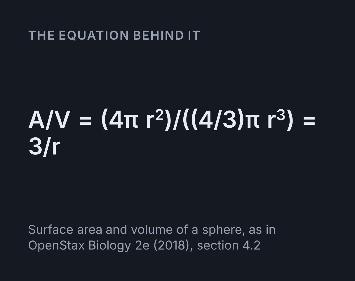
A cell takes in everything it needs, and gets rid of its wastes, through its membrane. For a
spherical cell of radius r the surface area is A = 4πr² and the volume
V = 4πr³/3, so the ratio is A/V = 3/r: as a cell grows, its
volume rises faster than its surface, and each cubic micrometre of cytoplasm is served by
less membrane. That is why cells stay small, divide, or change shape, which is the argument
OpenStax Biology makes in its section on cell size.
The slider beside the drawing sets a diameter from 1 to 100 µm and gives the area, the volume and the ratio. The shapes cells and organelles take to escape the limit are familiar ones: the folded inner membrane of a mitochondrion, the microvilli of a gut cell, the flat disc of a red blood cell.
Worked example: a 20 µm cell and a 40 µm cell
A spherical cell is 20 µm across. What is its surface area to volume ratio, and what happens when it doubles in diameter?
- The radius is 10 µm, so the surface area is 4π × 10² = 1,257 µm² and the volume is 4/3 × π × 10³ = 4,189 µm³.
- The ratio is 1,257 ÷ 4,189 = 0.3 per µm, which is 3/r with r = 10 µm.
- At 40 µm across the radius is 20 µm: the area is 5,027 µm² and the volume 33,510 µm³, four times the area and eight times the volume, so the ratio halves to 0.15 per µm.
- A bacterium 1 µm across has a ratio of 6 per µm, 20 times that of the 20 µm cell, which is one reason a bacterium manages without the internal membranes a larger cell relies on.
The explorer shows 0.3 per µm at its opening setting of 20 µm, and 0.15 per µm at 40 µm.
How big the parts are
The drawings are not to scale, because a ribosome drawn to scale in a 20 µm cell would be about a thousandth of its width. The typical sizes behind the size ladder are:
- Animal or plant cell: 10 to 100 µm
- Nucleus: 2 to 10 µm
- Red blood cell: 7 to 8 µm
- Chloroplast: 4 to 6 µm
- Bacterium: 100 nm to 5 µm
- Mitochondrion: 1 to 2 µm
- ER tubule: 30 to 100 nm
- Ribosome: 20 to 30 nm
- Cytoskeleton fibre: 7 to 25 nm
- Membrane thickness: 4 to 10 nm
A light microscope separates details down to about 200 nm, so it shows the nucleus, the cell wall, the vacuole and chloroplasts, and mitochondria as specks, but not ribosomes, membranes or the fibres of the cytoskeleton, which need an electron microscope. The Microscope Magnification Calculator works out how large a cell of a given size appears at a given magnification.
Sources: organelle sizes and the red blood cell from Milo and Phillips, Cell Biology by the Numbers (2015); cell sizes, the cytoskeleton, the microscope limit and every structure and function from OpenStax Biology 2e (Clark, Douglas and Choi, 2018), chapter 4.
What these diagrams do not show
- A real cell is three dimensional. Each drawing is a slice, and a mitochondrion that looks like an oval here may be part of a branched network.
- Every cell type is different. These are the typical cells textbooks describe. A mature red blood cell has no nucleus at all, a skeletal muscle cell has many, and cells of a root, which grow in the dark, generally have no chloroplasts.
- Other plastids are left out. Chloroplasts are one of several kinds of plastid, and plant cells also carry plastids that store starch or pigment.
- Numbers and positions are illustrative. A leaf mesophyll cell holds about 100 chloroplasts, and an active animal cell hundreds or thousands of mitochondria, far more than a clear diagram can show.
Common mistakes
- Saying plant cells have no mitochondria. They have both mitochondria and chloroplasts.
- Confusing the cell wall with the cell membrane. A plant cell has both: the membrane controls what enters, and the wall outside it gives strength and shape.
- Saying mitochondria make energy. They release energy stored in food and store it in ATP; energy is never made.
- Mixing up the two kinds of ER. The rough ER carries ribosomes and works on proteins; the smooth ER has few or none and makes lipids, detoxifies and stores calcium.
- Treating the nucleolus as an organelle with its own membrane. It is a dense region inside the nucleus, where ribosome subunits are assembled.
- Expecting to see ribosomes under a school microscope. At 20 to 30 nm they are far below what light can resolve.
Where to go next
Watch water cross a membrane in the Diffusion and Osmosis Simulator, follow a gene from the nucleus to a ribosome in the Protein Synthesis Simulator, see what limits a chloroplast’s output in the Photosynthesis Rate Simulator, and turn the molecule the nucleus holds in the DNA Double Helix Explorer.
Common questions
What is the difference between a plant cell and an animal cell?
A plant cell has a cellulose cell wall, chloroplasts and a large central vacuole, and an animal cell has none of them, while an animal cell has a centrosome and lysosomes that most plant cells lack. Everything else is shared: both have a cell membrane, cytoplasm, a nucleus, ribosomes, rough and smooth endoplasmic reticulum, a Golgi apparatus, mitochondria, peroxisomes and a cytoskeleton. The wall also gives a plant cell its fixed, often box-like shape, where an animal cell is rounder and more varied, and plant cells are joined to their neighbours through the wall by channels called plasmodesmata.
What are the main organelles of an animal cell and what do they do?
The nucleus holds the DNA and directs the cell; ribosomes build proteins; the rough endoplasmic reticulum folds and modifies those proteins and the smooth ER makes lipids and stores calcium; the Golgi apparatus sorts, packages and dispatches them in vesicles; mitochondria release the energy in food as ATP; lysosomes digest worn-out parts and engulfed bacteria; peroxisomes break down fatty acids and poisons; the centrosome organises microtubules; and the cell membrane controls what enters and leaves. The explorer gives the structure, the function and the size of each one.
Do plant cells have mitochondria?
Yes. Plant cells have mitochondria as well as chloroplasts, and the two do different jobs: chloroplasts use light to make sugar, and mitochondria release the energy in that sugar as ATP by aerobic respiration, just as they do in an animal cell. A plant respires all the time, in the dark as well as the light, and the cells of its roots, which receive no light, depend on their mitochondria entirely.
Why are cells so small?
Because a cell’s surface grows more slowly than its volume. For a sphere the surface area is 4πr² and the volume 4πr³/3, so the ratio is 3/r: a cell 20 µm across has 0.3 µm² of membrane for every cubic micrometre, and one 40 µm across only 0.15, half as much. Everything a cell uses and every waste it makes has to cross that membrane, so a cell that grew without limit would starve its own interior. Cells stay small, divide, or fold and flatten their membranes to keep the ratio up.
Which parts of a cell can you see with a light microscope?
The nucleus, the cell wall, the central vacuole and chloroplasts, and mitochondria only as small specks. A light microscope resolves details down to about 200 nm, OpenStax Biology’s figure, so ribosomes at 20 to 30 nm, cell membranes about 4 nm thick and the fibres of the cytoskeleton are all too small to resolve, and need an electron microscope. The explorer’s size ladder marks that 200 nm limit against the size of every part.