Heart Explorer
Free 3D heart anatomy: hide the wall to see the valves and papillary muscles, and trace the coronary arteries and cardiac veins by name. No sign-in.
Visualiser
Heart, 23 structures
Checking this device for a stored copy.
Start here
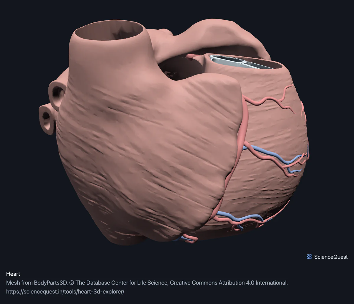
- Hide the Heart wall band This is the one thing to do first. The wall is a single mesh that encloses everything else, so with it shown you are looking at the outside of a heart. Hidden, the three valves, the five papillary muscles and the whole coronary tree are exposed in place.
- Click Coronary arteries twice, then Front Opening a band frames it, and this one starts open, so the second click frames the coronary tree. Front then shows it from the sternocostal surface, the face of the heart that lies behind the sternum and costal cartilages. The anterior interventricular artery runs down the groove between the ventricles.
- Turn on Cut plane and drag it Sections the wall rather than hiding it, so you can see the thickness of the left ventricle against the right. That difference in wall thickness is a measurement here, not an illustration.
Heart only. No aortic valve, no conducting system and no chamber meshes: the dataset publishes none of them. The great vessels beyond the coronary ostia belong to the cardiovascular region rather than this one, so the aorta and the pulmonary trunk are not here either.
- 23 structures
- 487k triangles
- 2500 KB, on request
- 0.000883 mm worst error per axis
- 1 heart wall
- 5 papillary muscles
- 3 valves
- 9 coronary arteries
- 5 cardiac veins
Where this comes from
Segmented from imaging of one adult male body, not drawn by an artist, so the shapes are measurements. Two consequences worth knowing: it is one person’s anatomy rather than an average, and faint stepping on some surfaces is the resolution of the original scan rather than a rendering fault.
Meshes from BodyParts3D, © The Database Center for Life Science, under Creative Commons Attribution 4.0 International. Names and identifiers are the dataset’s own: Foundational Model of Anatomy identifiers and English labels, as its parts list gives them. Converted meshes are shared under the same licence.
Changes made: Meshes were converted from binary STL to an indexed mesh with 16-bit quantised vertex positions, and surface normals were recomputed from the geometry. No vertex was moved other than by that quantisation, whose worst-case error on any one coordinate is reported on the page.
Latin terms for 9 of the 23 structures come from English Wikipedia anatomy infoboxes, under Creative Commons Attribution-ShareAlike 4.0, joined by Wikidata property P1402, Foundational Model of Anatomy identifier. Only a term a source states for that structure is used, never one constructed from a family name, which is why the rest carry no Latin rather than a guess.
All 23 structures
Every structure the model contains, group by group. Each name opens the viewer with that structure selected, so a link can point at one structure rather than at the whole region.
Heart wall 1
- wall of heart muscle
Papillary muscles 5
Valves 3
- tricuspid valve valvula tricuspidalis, valva atrioventricularis dextra other tissue
- mitral valve valva atrioventricularis sinistra, valva mitralis, valvula bicuspidalis other tissue
- pulmonary valve valva trunci pulmonalis other tissue
Coronary arteries 9
- stem of left coronary artery blood vessel
- trunk of right coronary artery blood vessel
- anterior interventricular branch of left coronary artery blood vessel
- circumflex branch of left coronary artery ramus circumflexus arteriae coronariae sinistrae blood vessel
- marginal branch of right coronary artery ramus marginalis dexter arteriae coronariae dextrae blood vessel
- posterior interventricular branch of right coronary artery blood vessel
- right posterolateral branch of right coronary artery blood vessel
- set of interventricular septal branches of left coronary artery blood vessel
- set of interventricular septal branches of right coronary artery blood vessel
Cardiac veins 5
- coronary sinus sinus coronarius blood vessel
- great cardiac vein vena cordis magna, vena cardiaca magna blood vessel
- middle cardiac vein vena cardiaca media, vena cordis media blood vessel
- set of anterior cardiac veins venae cardiacae anteriores, venae ventriculi dextri anteriores blood vessel
- set of posterior veins of left ventricle blood vessel
Citing this tool
Last updated . Add the date you accessed it as well, which a citation of a page that can change asks for. If a specific view matters, cite the page address as it stands in your browser instead: it records what is selected and shown.
Why hiding the wall is the whole tool
This 3D model of one adult human heart, from the BodyParts3D dataset, has its wall, the tricuspid, mitral and pulmonary valves, the papillary muscles and the coronary arteries and veins as separate structures to hide and label. The dataset treats a chamber as a region of the heart rather than as an object. There is no mesh called left ventricle; there is one large mesh called the wall of heart that carries the chamber shapes, and the structures filed under each chamber are its valve and its papillary muscles. So the heart arrives closed, and hiding one band opens it.
What is left is worth naming carefully. Three papillary muscles hang in the right ventricle, the anterior, posterior and septal, and two in the left. Their chordae are below the resolution of the segmentation and are not here, which is why the valves appear to float above the muscles that in life pull on them.
Three valves, not four
The tricuspid, mitral and pulmonary valves ship as separate meshes. The aortic valve does not. A heart has four valves, so this model is missing one, and the band is labelled for what it contains rather than for what a diagram would show. If you are checking this against a textbook, that is the gap you would find first, and you should find it acknowledged rather than papered over.
The coronary tree ends where the data ends
Both coronary arteries are here from their stems, with the anterior and posterior interventricular branches, the circumflex, the marginal, the right posterolateral and the septal branches on both sides. The venous side carries the coronary sinus and the great, middle, anterior and posterior cardiac veins.
What no segmentation can give you is the fine distal tree. Vessels thin continuously until they fall below the voxel size of the imaging, so each artery here stops at a cut end rather than tapering to nothing. That cut is an artefact of the method and not an anatomical fact, and it is the one thing on this model that would mislead if you read it as complete.
One person’s coronary anatomy
This is segmented from imaging of a single adult body, which matters more for a heart than for a femur. Coronary dominance, meaning which artery supplies the back of the heart, varies between people: the right artery does it in most, the left in roughly one in ten, and both share it in the rest. So the pattern here is that person’s pattern. It is a real heart rather than an idealised drawing, with exactly the caveat that comes with any single specimen.
Common questions
Why are there only three valves and not four?
Because the dataset publishes three. The tricuspid, mitral and pulmonary valves are here as separate meshes and the aortic valve is not, so rather than quietly listing three under a heading that claims the usual four, the band is labelled for what it holds. A heart has four valves. This model has three of them, and if you are checking the tool against a textbook that is the difference you would find first.
Where are the ventricles and atria?
In the wall. The dataset treats a chamber as a region of the heart rather than as an object, so there is no separate mesh named left ventricle: the chamber shape is part of one large mesh called the wall of heart, and the structures filed under each chamber are its valve and its papillary muscles. Hiding the wall is therefore what opens the heart up, and what is left is the valve ring, the papillary muscles hanging towards the apex and the coronary circulation.
Is the coronary tree complete?
It is complete as published, which is not the same as complete as it exists. Both coronary arteries are here with their named branches, the anterior and posterior interventricular, the circumflex, the marginal, the right posterolateral and the septal branches on both sides, along with the coronary sinus and the great, middle, anterior and posterior cardiac veins. What a segmentation cannot give you is the fine distal tree, which thins below the resolution of the imaging, so the arteries end where the data ends rather than where the vessel does.
Is this one person’s heart?
Yes, and that matters more for a heart than for a bone. It is segmented from imaging of a single adult body, so the coronary pattern you see is that person’s. Coronary anatomy is genuinely variable between people: which artery supplies the back of the heart is the common example, and it differs in roughly one person in five. So this is a real heart rather than an average or an idealised drawing, with the caveat that comes with any single specimen.