Hand and Wrist Skeleton Explorer
Turn all 27 bones of the hand and wrist in 3D, with the eight carpals separated so you can see their shapes rather than memorise a rhyme.
Visualiser
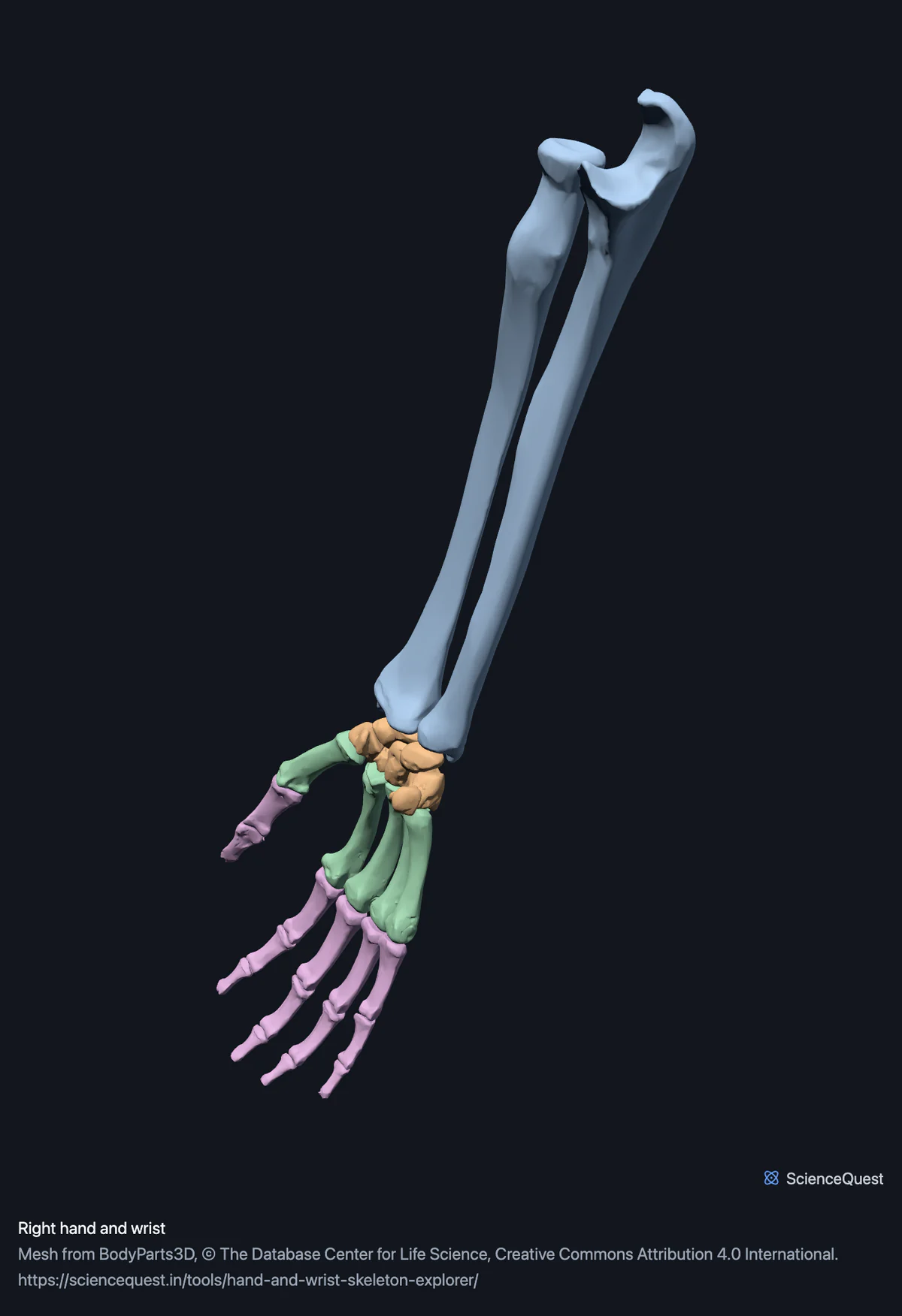
Right hand and wrist, 27 bones in 29 meshes
Checking this device for a stored copy.
Start here
- Select the scaphoid, then the lunate beside it The proximal row is curved so it can roll on the radius. That is the shape a mnemonic cannot give you.
- Hide the Forearm, Metacarpals and Phalanges groups Leaves the eight carpals alone, which is the only way to see how they interlock.
- Compare Radial and Ulnar The thumb is a separate column of bone set forward of the other four, and only a side view shows it.
Bone only. No ligaments, tendons, retinaculum or nerves, so this shows the skeleton the carpal tunnel passes through but not the tunnel. The radius and ulna are included whole rather than cut at the wrist.
- 27 bones
- 43k triangles
- 250 KB, on request
- 0.00305 mm worst error per axis
- 2 forearm
- 8 carpals
- 5 metacarpals
- 14 phalanges
The right radius and right ulna load with them for context and are not counted above. 29 meshes load in total, and every one of them is listed, nameable and selectable.
A hand has 27 bones: eight carpals, five metacarpals and fourteen phalanges. The radius and ulna load with them because the wrist joint is the articulation between the radius and the proximal carpal row, so 29 meshes arrive and 27 of them are bones of the hand.
Where this comes from
Segmented from imaging of one adult male body, not drawn by an artist, so the shapes are measurements. Two consequences worth knowing: it is one person’s anatomy rather than an average, and faint stepping on some surfaces is the resolution of the original scan rather than a rendering fault.
Meshes from BodyParts3D, © The Database Center for Life Science, under Creative Commons Attribution 4.0 International. Names and identifiers are the dataset’s own: Foundational Model of Anatomy identifiers and English labels, as its parts list gives them. Converted meshes are shared under the same licence.
Changes made: Meshes were converted from binary STL to an indexed mesh with 16-bit quantised vertex positions, and surface normals were recomputed from the geometry. No vertex was moved other than by that quantisation, whose worst-case error on any one coordinate is reported on the page.
Latin terms for 6 of the 29 structures come from English Wikipedia anatomy infoboxes, under Creative Commons Attribution-ShareAlike 4.0, joined by Wikidata property P1402, Foundational Model of Anatomy identifier. Only a term a source states for that structure is used, never one constructed from a family name, which is why the rest carry no Latin rather than a guess.
All 29 structures
Every structure the model contains, group by group. Of these, 27 are bones of this region. Each name opens the viewer with that structure selected, so a link can point at one structure rather than at the whole region.
Forearm 2
- right radius radius
- right ulna ulna
Carpals 8
- right scaphoid
- right lunate
- right triquetral os triquetrum, os pyramidale
- right pisiform os pisiforme
- right trapezium os trapezium, os multangulum majus
- right trapezoid
- right capitate
- right hamate os hamatum
Metacarpals 5
Phalanges 14
- proximal phalanx of right thumb
- distal phalanx of right thumb
- proximal phalanx of right index finger
- middle phalanx of right index finger
- distal phalanx of right index finger
- proximal phalanx of right middle finger
- middle phalanx of right middle finger
- distal phalanx of right middle finger
- proximal phalanx of right ring finger
- middle phalanx of right ring finger
- distal phalanx of right ring finger
- proximal phalanx of right little finger
- middle phalanx of right little finger
- distal phalanx of right little finger
Citing this tool
Last updated . Add the date you accessed it as well, which a citation of a page that can change asks for. If a specific view matters, cite the page address as it stands in your browser instead: it records what is selected and shown.
Eight carpals, two rows
Radial side first, which is the thumb side, in both rows:
- Proximal row: scaphoid, lunate, triquetral, pisiform.
- Distal row: trapezium, trapezoid, capitate, hamate.
The trapezium and the trapezoid are the pair people confuse. Keep them apart by what they carry rather than by their names: the trapezium takes the thumb, the trapezoid sits under the index finger.
The words on the view buttons
Medial and lateral are ambiguous for a hand, because the forearm rotates and they only mean anything if you assume the anatomical position. These do not:
- Radial
- The thumb side, whatever the arm is doing.
- Ulnar
- The little finger side.
- Palmar
- The palm.
- Dorsal
- The back of the hand.
Common questions
What order are the carpal bones in?
Proximal row from the thumb side: scaphoid, lunate, triquetral, pisiform. Distal row in the same direction: trapezium, trapezoid, capitate, hamate. The mnemonics for that order are fine, but they teach you a sequence rather than a set of shapes, which is why the sequence tends to survive an exam and not much longer. Isolating each bone and turning it is the part a rhyme cannot do: the proximal row is curved so it can roll on the radius, and once you have seen that the order stops being arbitrary.
What is the difference between the trapezium and the trapezoid?
They are different bones and the names are unhelpfully similar. The trapezium is the one that carries the thumb, and its saddle-shaped joint is what allows opposition, which is most of what makes a human hand useful. The trapezoid sits immediately next to it, under the index finger, and does very little by comparison. The reliable way to keep them apart is not the names but the relationship: select each in turn and see which metacarpal it meets.
Why does a scaphoid fracture matter more than other wrist fractures?
Because of how its blood supply runs. The scaphoid is the most commonly fractured carpal, and its vessels enter towards the distal end and travel backwards through the bone. A fracture across the waist can therefore leave the proximal fragment with no supply at all, which risks the bone dying rather than healing. Isolating it in the model shows why there is so little room for vessels: most of its surface is articular, so there are few places for anything to enter.
Why do the view buttons say radial and ulnar instead of medial and lateral?
Because the forearm rotates. Medial and lateral only mean something for a hand if you assume the anatomical position, palm forwards, and nobody holds their hand that way. The radial side is the thumb side whatever the arm is doing, so radial and ulnar stay unambiguous. Front and back are palmar and dorsal for the same reason. It is a small thing, but using the wrong pair is the sort of habit that has to be unlearned later.