Male Reproductive Organs Explorer
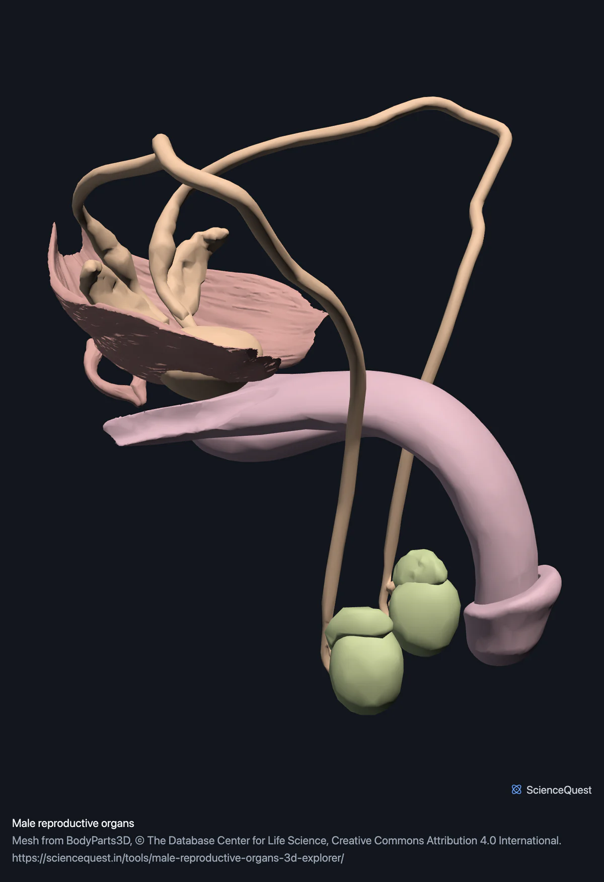
Turn the male reproductive organs in 3D: testes, epididymides, deferent ducts, seminal vesicles and prostate, with the pelvic floor around the prostate.
Visualiser
Male reproductive organs, 15 structures
Checking this device for a stored copy.
Start here
- Hide the Pelvic floor muscles They are two thirds of the geometry here and they wrap round the prostate and seminal vesicles, so hiding them is what turns a solid block into a set of separable organs.
- Click Ducts, seminal vesicles and prostate twice, then Left Opening a band frames it, and this one starts open, so the second click is the one that takes you there. The side view is the one that reads. It shows the deferent duct arching over from the testis, the seminal vesicle behind, and the prostate below, which is the order semen travels in.
- Open the internal organs model in another tab The bladder and ureters are there, not here, and both models share one coordinate frame. Together they show the prostate sitting directly beneath the bladder.
Organs, and the pelvic floor muscles around the prostate. No female anatomy, because the specimen is male. No bladder and no kidneys: those are in the internal organs model. The urethra is not in any model on this site. No prostatic zones, no seminiferous tubules and no spermatic cord as a separate structure.
- 15 structures
- 71k triangles
- 400 KB, on request
- 0.00134 mm worst error per axis
- 4 testes and epididymides
- 5 ducts, seminal vesicles and prostate
- 3 penis
- 3 pelvic floor muscles
Where this comes from
Segmented from imaging of one adult male body, not drawn by an artist, so the shapes are measurements. Two consequences worth knowing: it is one person’s anatomy rather than an average, and faint stepping on some surfaces is the resolution of the original scan rather than a rendering fault.
Meshes from BodyParts3D, © The Database Center for Life Science, under Creative Commons Attribution 4.0 International. Names and identifiers are the dataset’s own: Foundational Model of Anatomy identifiers and English labels, as its parts list gives them. Converted meshes are shared under the same licence.
Changes made: Meshes were converted from binary STL to an indexed mesh with 16-bit quantised vertex positions, and surface normals were recomputed from the geometry. No vertex was moved other than by that quantisation, whose worst-case error on any one coordinate is reported on the page.
Latin terms for 10 of the 15 structures come from English Wikipedia anatomy infoboxes, under Creative Commons Attribution-ShareAlike 4.0, joined by Wikidata property P1402, Foundational Model of Anatomy identifier. Only a term a source states for that structure is used, never one constructed from a family name, which is why the rest carry no Latin rather than a guess.
All 15 structures
Every structure the model contains, group by group. Each name opens the viewer with that structure selected, so a link can point at one structure rather than at the whole region.
Testes and epididymides 4
- right testis testis organ
- left testis testis organ
- right epididymis epididymis organ
- left epididymis epididymis organ
Ducts, seminal vesicles and prostate 5
- right deferent duct organ
- left deferent duct organ
- right seminal vesicle vesicula seminalis, glandula vesiculosa organ
- left seminal vesicle vesicula seminalis, glandula vesiculosa organ
- prostate prostata organ
Penis 3
- corpus cavernosum of penis corpus cavernosum penis organ
- corpus spongiosum of penis organ
- glans penis glans penis organ
Pelvic floor muscles 3
- right pubococcygeus muscle
- left pubococcygeus muscle
- external anal sphincter sphincter ani externus muscle
Citing this tool
Last updated . Add the date you accessed it as well, which a citation of a page that can change asks for. If a specific view matters, cite the page address as it stands in your browser instead: it records what is selected and shown.
One body, and it is male
Every model on this site is segmented from the same single adult body. That is what lets the skeleton, the organs, the brain and the vessels share one coordinate frame, so a prostate here and a pelvis there are the same millimetres apart as they are in a person rather than being two drawings at two scales.
The cost of that is direct: anything absent from that one body is absent from every model. The body is male, so there is no female reproductive anatomy to show, and no amount of work on this site can add it from this dataset. It is a limit of the source rather than a decision made here, and you should know which it is.
Why muscles are in a reproductive model
Both pubococcygei and the external anal sphincter are filed here by the dataset, and they are roughly two thirds of the geometry. They are also the reason the rest makes sense: the pelvic floor is what the organs rest on, and stripping it out would leave a prostate and two seminal vesicles floating with nothing to locate them against.
They are tagged as muscle rather than as organs, so the model reports 12 organs and 3 muscle structures. Counting all 15 as organs would have been the easy inaccuracy.
What the resolution gives you, and what it does not
Position, clearly. The prostate lies directly in front of the rectum, which is why it can be felt through the rectal wall. The rectum is in the internal organs model rather than this one, and the two share one coordinate frame, so opened together they show that relationship as a measurement rather than as a schematic. The prostate also wraps the first stretch of the urethra just below the bladder, which you have to take on trust here: the urethra is not in any model on this site. The deferent duct’s course from the epididymis up and over into the pelvis is visible in full, which is hard to follow in any flat illustration.
Internal structure, not at all. The prostate is one smooth surface of 1,754 triangles with no zones, so the peripheral, central and transition zones that matter clinically are not here. Nor are the seminiferous tubules inside the testis, and the spermatic cord is not a separate structure: what you get is the duct within it.
Common questions
Why is there no female anatomy?
Because BodyParts3D is one adult male body, so there is no female reproductive anatomy in the dataset to show. That is a limit of the source rather than a decision made here, and it is worth stating plainly: every model on this site comes from the same single specimen, which is what makes the regions share one coordinate frame, and the cost of that is that anything absent from that body is absent everywhere.
Why are pelvic floor muscles in a reproductive model?
Because the dataset files them here and because they are most of what you are looking at. Both pubococcygei and the external anal sphincter are about two thirds of this region’s geometry, and they are the floor the organs rest on, so removing them would leave the prostate and seminal vesicles floating with nothing to place them against. They are tagged as muscle rather than as organs, so the model honestly reports 12 organs and 3 muscle structures.
Is the prostate anatomically useful at this resolution?
For position, yes. For internal structure, no. The prostate here is a single smooth surface of under two thousand triangles, so you can see exactly where it sits on the pelvic floor. It lies directly in front of the rectum, which is why it can be felt through the rectal wall, and it surrounds the first stretch of the urethra below the bladder, though neither the rectum nor the urethra is in this model. What you cannot see are its zones, the peripheral, central and transition zones that matter clinically, because the segmentation did not divide it.
How does this relate to the urinary organs?
They are in the internal organs model, and the two are designed to be opened together. The bladder, both ureters and the kidneys ship there, this region carries the prostate and the ducts, and because every region shares one coordinate frame the prostate sits in its real relationship to the bladder above it. The urethra is not in any model here: the dataset publishes it, but neither this region nor the internal organs model ships it.