Testis
The testes are the male gonads, a pair of oval organs in the scrotum. They make sperm and androgens such as testosterone. (OpenStax A&P 2e, 27.1; StatPearls, Abdomen and Pelvis: Testes)
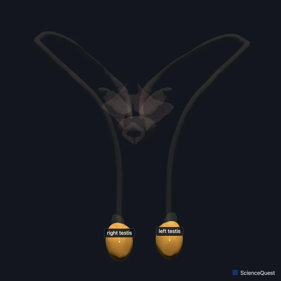
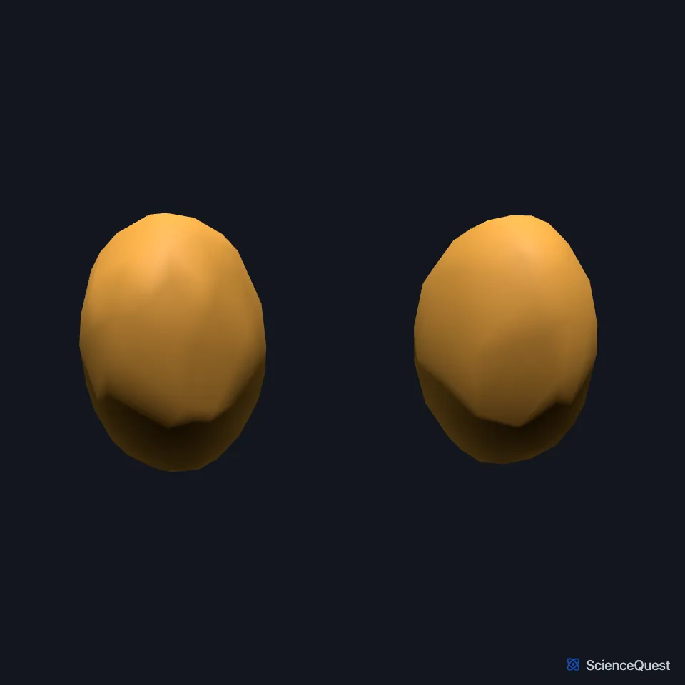
Open it in the Male Reproductive Organs Explorer See it alone in 3D
Where are the testes?
They hang in the scrotum on either side of the scrotal septum, and the left is usually a little lower than the right. The spermatic cord suspends each testis from above, and the scrotal ligament, a remnant of the gubernaculum, ties its lower end to the scrotum. (StatPearls, Abdomen and Pelvis: Testes)
The scrotum holds the testes a few degrees below core body temperature, which sperm production needs. The cremaster muscles raise or lower the testes from day to day as the temperature changes. (OpenStax A&P 2e, 27.1; StatPearls, Abdomen and Pelvis, Scrotum and Abdomen and Pelvis: Testes)
Each testis forms in the abdomen and descends through the abdominal wall and inguinal canal into the scrotum, in the seventh month of fetal life. (OpenStax A&P 2e, 27.1; StatPearls, Abdomen and Pelvis: Testes)
What is a testis made of?
Its outer coverings are the tunica vaginalis, a sac with parietal and visceral layers, and beneath it the tough white tunica albuginea; Gray’s 1918 text adds a third, innermost tunic, the tunica vasculosa. The tunica albuginea turns inward along the back of the testis, and septa radiate from there to split the testis into cone-shaped lobules. OpenStax counts 300 to 400 lobules, while Gray’s 1918 text cites older estimates of 250 and 400. (OpenStax A&P 2e, 27.1; StatPearls, Abdomen and Pelvis: Testes; Gray 1918, 3c. The Male Genital Organs)
Tightly coiled seminiferous tubules fill each lobule. Sperm pass from them into the straight tubules and the rete testis, then leave through the efferent ductules, 15 to 20 of them in OpenStax and 12 to 20 in Gray’s 1918 text. (OpenStax A&P 2e, 27.1; Gray 1918, 3c. The Male Genital Organs)
What does the testis do?
Sperm develop in the seminiferous tubules from puberty on, starting from germ cells near the tubule wall. New cycles start at intervals, and the tubules do not all keep in step. (OpenStax A&P 2e, 27.1; StatPearls, Physiology, Male Reproductive System)
Sertoli cells reach from the base of the tubule wall to its lumen and nurse the developing sperm, controlling the ions and amino acids around them. Tight junctions between neighboring Sertoli cells form the blood-testis barrier, which hides the later germ cells from the immune system. (OpenStax A&P 2e, 27.1; StatPearls, Histology, Sertoli Cell)
Leydig cells, between the tubules, make testosterone, about 6 to 7 mg a day. Luteinizing hormone from the anterior pituitary gland controls testosterone output, and follicle-stimulating hormone controls sperm production. (OpenStax A&P 2e, 27.1; StatPearls, Abdomen and Pelvis: Testes)
What lies next to the testis?
The epididymis runs along the back of each testis, with its head at the upper pole. The tunica vaginalis covers the testis except at the back and top, where the epididymis and the spermatic cord attach. (StatPearls, Abdomen and Pelvis: Testes)
The testicular arteries leave the abdominal aorta just below the renal arteries, cross over the ureter and reach the testis in the spermatic cord. The artery of the deferent duct and the cremasteric artery add to the supply. (StatPearls, Abdomen and Pelvis: Testes)
Its veins form the pampiniform plexus, which joins into the testicular vein. The right testicular vein drains into the inferior vena cava and the left into the left renal vein, and lymph follows the arteries to the preaortic nodes. (StatPearls, Abdomen and Pelvis: Testes)
Where the sources differ
OpenStax and Holstein and colleagues (2003) say a new cycle of sperm production starts about every 16 days; StatPearls says about every 13 days. OpenStax and StatPearls both say the cycles are not in step across the tubules. (OpenStax A&P 2e, 27.1; Holstein et al. 2003, Reprod Biol Endocrinol; StatPearls, Physiology, Male Reproductive System)
The sources give different times for making a sperm: OpenStax gives about 64 days from spermatogonium to formed sperm, while Holstein and colleagues (2003) give 4.6 cycles, or 74 days, in the testis. (OpenStax A&P 2e, 27.1; Holstein et al. 2003, Reprod Biol Endocrinol)
OpenStax gives each adult testis as about 4 to 5 cm long. StatPearls gives 3 to 5 cm long and 2 to 3 cm wide. (OpenStax A&P 2e, 27.1; StatPearls, Abdomen and Pelvis: Testes)
| Feature | Detail and source |
|---|---|
| Length | About 4 to 5 cm (OpenStax); 3 to 5 cm (StatPearls) (OpenStax A&P 2e, 27.1; StatPearls, Abdomen and Pelvis: Testes) |
| Coverings | Tunica vaginalis, then tunica albuginea (Gray 1918 adds the tunica vasculosa) (OpenStax A&P 2e, 27.1; Gray 1918, 3c. The Male Genital Organs) |
| Lobules | 300 to 400 (OpenStax); 250 or 400, two older estimates cited by Gray 1918 (OpenStax A&P 2e, 27.1; Gray 1918, 3c. The Male Genital Organs) |
| Artery | Testicular artery, from the abdominal aorta (StatPearls, Abdomen and Pelvis: Testes) |
Touches in this model
Measured on the BodyParts3D meshes: surfaces within 2 mm of the testis, nearest first.
- Left deferent duct in contact
- Left epididymis in contact
- Right deferent duct in contact
- Right epididymis in contact
Sources
- OpenStax Anatomy and Physiology 2e, section 27.1, Anatomy and Physiology of the Testicular Reproductive System. CC BY-NC-SA 4.0. Read 2026-10-25.
- StatPearls, Anatomy, Abdomen and Pelvis: Testes. Manpreet S. Tiwana; Stephen W. Leslie. Last update April 15, 2023. CC BY-NC-ND 4.0. Read 2026-10-25.
- StatPearls, Anatomy, Abdomen and Pelvis, Scrotum. Rosa A. Garcia; Hussain Sajjad. Last update July 24, 2023. CC BY-NC-ND 4.0. Read 2026-10-25.
- Gray’s Anatomy of the Human Body, 20th US edition (1918), XI. Splanchnology, 3c. The Male Genital Organs, 1. The Testes and their Coverings (1918 terms: tubuli seminiferi, tubuli recti and ductuli efferentes for seminiferous tubules, straight tubules and efferent ductules). Henry Gray. Public domain. Read 2026-10-25.
- StatPearls, Physiology, Male Reproductive System. Purnima Gurung; Ekrem Yetiskul; Ishwarlal Jialal. Last update May 1, 2023. CC BY-NC-ND 4.0. Read 2026-10-25.
- StatPearls, Histology, Sertoli Cell. Wah J. Wong; Yusuf S. Khan. Last update November 14, 2022. CC BY-NC-ND 4.0. Read 2026-10-25.
- Reproductive Biology and Endocrinology, Understanding spermatogenesis is a prerequisite for treatment, Reprod Biol Endocrinol 1, article 107 (full text, PMC293421; PMID 14617369). Holstein AF, Schulze W, Davidoff M. BioMed Central open access; verbatim copying and redistribution permitted with the notice. Read 2026-10-25.
The values are facts and free to use; this page’s selection and presentation are © 2026 ScienceQuest.