Great Vessels Explorer
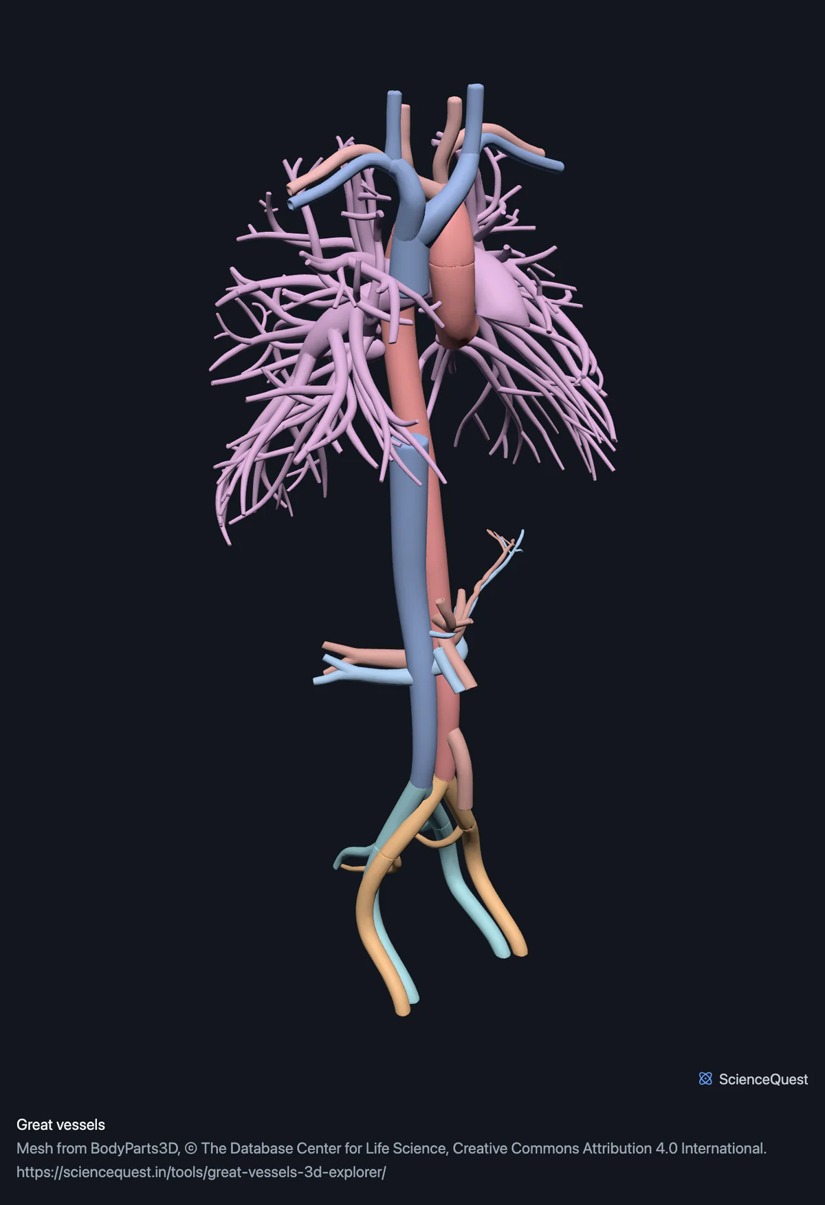
Turn the great vessels in 3D: the aorta and its named branches, both venae cavae, the pulmonary vessels and the renal, carotid and iliac vessels.
Visualiser
Great vessels, 42 structures
Checking this device for a stored copy.
Start here
- Hide the two Pulmonary vessels They are 70 percent of the model’s triangles on their own, because the segmentation followed them deep into both lungs. Hidden, the aorta and the venae cavae are no longer buried inside a branching thicket.
- Click Aorta, then Left The arch and its three branches read properly only from the side. From the front the ascending and descending aorta overlap each other almost exactly.
- Show only Abdominal arteries and Abdominal veins The clearest way to see how the coeliac trunk, the mesenteric arteries and the renal vessels are stacked on the aorta, and how the left renal vein has to cross the midline in front of it.
Blood vessels only. Nothing distal to the first named branches: no vessels in the limbs, none in the head beyond the carotids, and no capillary bed. The coronary arteries and cardiac veins are in the heart model, not here.
- 42 structures
- 270k triangles
- 1500 KB, on request
- 0.00432 mm worst error per axis
- 3 aorta
- 5 arteries to head and arms
- 8 abdominal arteries
- 6 pelvic arteries
- 2 pulmonary vessels
- 8 venae cavae and tributaries
- 4 abdominal veins
- 6 pelvic veins
Where this comes from
Segmented from imaging of one adult male body, not drawn by an artist, so the shapes are measurements. Two consequences worth knowing: it is one person’s anatomy rather than an average, and faint stepping on some surfaces is the resolution of the original scan rather than a rendering fault.
Meshes from BodyParts3D, © The Database Center for Life Science, under Creative Commons Attribution 4.0 International. Names and identifiers are the dataset’s own: Foundational Model of Anatomy identifiers and English labels, as its parts list gives them. Converted meshes are shared under the same licence.
Changes made: Meshes were converted from binary STL to an indexed mesh with 16-bit quantised vertex positions, and surface normals were recomputed from the geometry. No vertex was moved other than by that quantisation, whose worst-case error on any one coordinate is reported on the page.
Latin terms for 39 of the 42 structures come from English Wikipedia anatomy infoboxes, under Creative Commons Attribution-ShareAlike 4.0, joined by Wikidata property P1402, Foundational Model of Anatomy identifier. Only a term a source states for that structure is used, never one constructed from a family name, which is why the rest carry no Latin rather than a guess.
All 42 structures
Every structure the model contains, group by group. Each name opens the viewer with that structure selected, so a link can point at one structure rather than at the whole region.
Aorta 3
- ascending aorta aorta ascendens, pars ascendens aortae blood vessel
- arch of aorta arcus aortae blood vessel
- descending aorta aorta descendens, pars descendens aortae blood vessel
Arteries to head and arms 5
- brachiocephalic artery blood vessel
- right common carotid artery arteria carotis communis blood vessel
- left common carotid artery arteria carotis communis blood vessel
- right subclavian artery arteria subclavia blood vessel
- left subclavian artery arteria subclavia blood vessel
Abdominal arteries 8
- celiac artery truncus coeliacus, arteria coeliaca blood vessel
- common hepatic artery arteria hepatica communis blood vessel
- left gastric artery arteria gastrica sinistra blood vessel
- splenic artery arteria splenica, arteria lienalis blood vessel
- superior mesenteric artery arteria mesenterica superior blood vessel
- inferior mesenteric artery arteria mesenterica inferior blood vessel
- right renal artery arteria renalis blood vessel
- left renal artery arteria renalis blood vessel
Pelvic arteries 6
- right common iliac artery arteria iliaca communis blood vessel
- left common iliac artery arteria iliaca communis blood vessel
- right external iliac artery arteria iliaca externa blood vessel
- left external iliac artery arteria iliaca externa blood vessel
- right internal iliac artery arteria iliaca interna blood vessel
- left internal iliac artery arteria iliaca interna blood vessel
Pulmonary vessels 2
- pulmonary artery arteria pulmonalis blood vessel
- pulmonary vein venae pulmonales blood vessel
Venae cavae and tributaries 8
- superior vena cava vena cava superior, vena maxima blood vessel
- inferior vena cava vena cava inferior blood vessel
- right brachiocephalic vein vena brachiocephalica, vena anonyma blood vessel
- left brachiocephalic vein vena brachiocephalica, vena anonyma blood vessel
- right internal jugular vein vena jugularis interna blood vessel
- left internal jugular vein vena jugularis interna blood vessel
- right subclavian vein vena subclavia blood vessel
- left subclavian vein vena subclavia blood vessel
Abdominal veins 4
- right renal vein blood vessel
- left renal vein blood vessel
- splenic vein vena lienalis blood vessel
- superior mesenteric vein vena mesenterica superior blood vessel
Pelvic veins 6
- right common iliac vein vena iliaca communis blood vessel
- left common iliac vein vena iliaca communis blood vessel
- right external iliac vein vena iliaca externa blood vessel
- left external iliac vein vena iliaca externa blood vessel
- right internal iliac vein vena iliaca interna, vena hypogastrica blood vessel
- left internal iliac vein vena iliaca interna, vena hypogastrica blood vessel
Citing this tool
Last updated . Add the date you accessed it as well, which a citation of a page that can change asks for. If a specific view matters, cite the page address as it stands in your browser instead: it records what is selected and shown.
Defined by subtraction, and the subtraction is enforced
This region is the cardiovascular meshes that are not part of the heart. The coronary arteries, the cardiac veins and the valves ship in the heart model, so sending them here as well would mean two copies of the same vessel that could quietly drift apart.
That definition is checked rather than described. The generator takes the leaves under the cardiovascular system, subtracts the leaves under the heart, and asserts the result equals this part list exactly, in both directions. A vessel cannot end up in both models, and it cannot end up in neither.
How far each vessel goes is not uniform
The pulmonary artery mesh alone is 43 percent of this model’s triangles and the pulmonary vein 27 percent, while the entire aorta is 11 percent. That is not a fact about calibre: the pulmonary vessels were segmented down into their branching trees inside both lungs, and the aorta was published as three smooth segments. Together the two pulmonary meshes are about 70 percent of the model.
Everywhere else the tree stops at the first named branch. You get the coeliac trunk with its hepatic, splenic and left gastric branches, both renals, both mesenterics and the iliacs down to internal and external. Nothing goes into a limb. If you are using this to trace a supply, it will take you to the organ and then stop.
The left gastric artery arrives without its right counterpart
A right gastric artery does exist. It is a small branch of the hepatic artery running along the lesser curvature of the stomach to meet the left, and BodyParts3D does not publish it, which is why only the left is here. Every other region is checked by a rule that a structure named for one side must have its mirror, and that rule is what would catch a skull shipped with a left parietal bone and no right one. Vessels break it legitimately, because vascular naming is genuinely asymmetric: there is no right counterpart to the brachiocephalic artery or to the coronary sinus. So this region and the heart are the two exemptions, and both are named in the test with the reason rather than switched off quietly.
What the relationships are good for
The vessels are in the same coordinate frame as every other model here, which is what makes them worth turning rather than reading about. The left renal vein crossing in front of the aorta, the arch passing over the left main bronchus, the bifurcation sitting at the level of the fourth lumbar vertebra: those are measured positions from one body, so they can be checked against the skeleton and the organ models rather than taken on trust from a drawing.
Common questions
Why are the coronary arteries not in here?
Because they are in the heart model, and shipping them twice would mean two versions of the same vessel that could drift apart. This region is defined as the cardiovascular meshes that are not part of the heart, and that definition is enforced rather than described: the build asserts this part list equals exactly the difference between the two, so a vessel cannot be in both and cannot be in neither.
How far do the vessels go?
To the first named branch and rarely further. You get the aorta in three parts, the brachiocephalic artery, both carotids and subclavians, the coeliac trunk with its hepatic, splenic and left gastric branches, both renal arteries, both mesenteric arteries, and the iliacs down to internal and external on each side. On the venous side both venae cavae, the brachiocephalic, jugular and subclavian veins, the renal and splenic veins, the superior mesenteric vein and the iliacs. What is not here is everything distal to those: no vessels in the limbs, none in the head beyond the carotid, and no capillary bed anywhere.
Why is there a left gastric artery and no right one?
Because this dataset does not publish the right one, not because it does not exist. A right gastric artery is a real vessel: a small branch of the hepatic artery that runs along the lesser curvature of the stomach to anastomose with the left. BodyParts3D has no mesh for it, so only the left is here. Vascular naming is genuinely asymmetric in other places, though, which is the reason this region is exempt from the check that otherwise requires every left structure to have a right one: nothing is called the right brachiocephalic artery, and the coronary sinus, a vein, has no right counterpart. That check is what catches a missing left parietal bone in the skull, so it is kept strict everywhere it is meaningful and switched off, by name and with a reason, where it is not.
Why is the pulmonary artery so much bigger than the aorta?
In the mesh, not in life. The pulmonary artery mesh has four times as many triangles as the aorta’s three segments together, because the pulmonary mesh includes the branching tree down into both lungs while the aorta is published as three smooth segments. It is a difference in how far the segmentation followed each vessel, not a difference in the vessels, and it is worth knowing before you compare their calibres by eye.