Rib Cage and Thoracic Spine Explorer
Turn the rib cage in 3D with all 24 ribs separable, the twelve thoracic vertebrae they hinge on, and the sternum in its three real pieces.
Visualiser
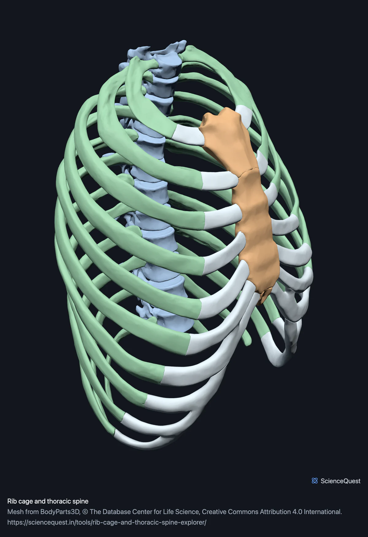
Rib cage and thoracic spine, 39 bones in 55 meshes
Checking this device for a stored copy.
Start here
- Turn to Front and follow the eleventh and twelfth ribs forward They stop short and reach nothing, which is what floating means. The upper seven curve round towards the sternum.
- Hide the Ribs group Leaves the column and the sternum, so the facets each rib hinges on are visible along the vertebral bodies.
- Select the manubrium and turn to Left Its joint with the body of the sternum sits level with the second rib, which is how ribs are counted on a patient.
Bone and costal cartilage. All sixteen costal cartilage meshes the dataset publishes are here, in their own pale band. They were left out at first and the cage looked broken rather than incomplete: every rib ended in mid air a long way from the sternum. No sternoclavicular joint, no intervertebral discs, no diaphragm and no lungs.
- 39 bones
- 990k triangles
- 5400 KB, on request
- 0.00293 mm worst error per axis
- 12 thoracic vertebrae
- 3 sternum
- 24 ribs
- 16 costal cartilage
The model also carries 16 of cartilage, so 55 parts load in total. They are listed and coloured like the rest, just not counted as bone.
39 bones, within 55 meshes. A thoracic cage is usually counted as 37: 24 ribs, 12 thoracic vertebrae and one sternum. The sternum is here as its three parts, manubrium, body and xiphoid process, which adds two. The remaining 16 meshes are costal cartilages and are not bone.
Where this comes from
Segmented from imaging of one adult male body, not drawn by an artist, so the shapes are measurements. Two consequences worth knowing: it is one person’s anatomy rather than an average, and faint stepping on some surfaces is the resolution of the original scan rather than a rendering fault.
Meshes from BodyParts3D, © The Database Center for Life Science, under Creative Commons Attribution 4.0 International. Names and identifiers are the dataset’s own: Foundational Model of Anatomy identifiers and English labels, as its parts list gives them. Converted meshes are shared under the same licence.
Changes made: Meshes were converted from binary STL to an indexed mesh with 16-bit quantised vertex positions, and surface normals were recomputed from the geometry. No vertex was moved other than by that quantisation, whose worst-case error on any one coordinate is reported on the page.
Latin terms for 1 of the 55 structures come from English Wikipedia anatomy infoboxes, under Creative Commons Attribution-ShareAlike 4.0, joined by Wikidata property P1402, Foundational Model of Anatomy identifier. Only a term a source states for that structure is used, never one constructed from a family name, which is why the rest carry no Latin rather than a guess.
All 55 structures
Every structure the model contains, group by group. Of these, 39 are bones of this region. Each name opens the viewer with that structure selected, so a link can point at one structure rather than at the whole region.
Thoracic vertebrae 12
Sternum 3
- manubrium
- body of sternum
- xiphoid process processus xiphoideus
Ribs 24
- right first rib
- left first rib
- right second rib
- left second rib
- right third rib
- left third rib
- right fourth rib
- left fourth rib
- right fifth rib
- left fifth rib
- right sixth rib
- left sixth rib
- right seventh rib
- left seventh rib
- right eighth rib
- left eighth rib
- right ninth rib
- left ninth rib
- right tenth rib
- left tenth rib
- right eleventh rib
- left eleventh rib
- right twelfth rib
- left twelfth rib
Costal cartilage 16
- right first costal cartilage cartilage
- left first costal cartilage cartilage
- right second costal cartilage cartilage
- left second costal cartilage cartilage
- right third costal cartilage cartilage
- left third costal cartilage cartilage
- right fourth costal cartilage cartilage
- left fourth costal cartilage cartilage
- right fifth costal cartilage cartilage
- left fifth costal cartilage cartilage
- right sixth costal cartilage cartilage
- left sixth costal cartilage cartilage
- right seventh costal cartilage cartilage
- left seventh costal cartilage cartilage
- right costal cartilage cartilage
- left costal cartilage cartilage
Citing this tool
Last updated . Add the date you accessed it as well, which a citation of a page that can change asks for. If a specific view matters, cite the page address as it stands in your browser instead: it records what is selected and shown.
True, false and floating
A grouping by what a rib reaches at the front, not by the bone itself:
- True, ribs one to seven
- Reach the sternum through their own costal cartilage.
- False, ribs eight to ten
- Join the cartilage of the rib above rather than the sternum directly.
- Floating, ribs eleven and twelve
- End in the abdominal wall and reach nothing at the front.
A sternum in three pieces
The manubrium, the body and the xiphoid process ossify separately, and the xiphoid usually fuses to the body by middle age, while the manubrium usually stays joined to the body by cartilage for life. The dataset keeps all three apart. Measured here, the manubrium sits about 140 mm above the xiphoid, so the sternum is longer than most diagrams imply.
Common questions
What are true, false and floating ribs?
A grouping by what the rib reaches at the front, not by the bone itself. The first seven pairs join the sternum through their own costal cartilage and are called true ribs. The eighth, ninth and tenth join the cartilage above them rather than the sternum directly, which makes them false ribs. The eleventh and twelfth end in the muscle of the abdominal wall and reach nothing at the front at all, which is why they are called floating. This model carries the bone, so the distinction shows up as which ribs curve round towards the sternum and which stop short: turn to the front view and follow the eleventh and twelfth forward to see them end.
Why does the sternum come apart into three pieces?
Because its three parts ossify separately, and the dataset segments them apart. The xiphoid usually fuses to the body by middle age, but the manubrium usually stays joined to the body by cartilage for life. The manubrium is the broad top piece that meets the clavicles, the body is the long middle, and the xiphoid process is the small tip, which is cartilage in youth and often does not fully ossify until middle age. Measured on this specimen the manubrium sits about 140 millimetres above the xiphoid, so the sternum is a good deal longer than most diagrams suggest. The joint between the manubrium and the body is a real landmark: it sits level with the second rib, which is how you count ribs on a patient.
Why are the ribs so much heavier to download than the skull?
Because of their shape. The 24 ribs are 755,704 triangles between them, averaging about 31,500 each, and that total is three times the whole skull. A rib is a long thin curved shell, and that is the worst case for a triangle budget: a surface that encloses almost no volume has nothing to amortise its own area against, where a compact bone like a vertebra gets a lot of shape for comparatively few triangles. That makes this the heaviest skeletal region on the site after the full skeleton, and the size is printed on the load button so you can decide before it downloads.
Are the costal cartilages included?
Yes, all sixteen costal cartilage meshes the dataset publishes, in their own pale band. This was the first skeletal region here to carry anything other than bone, and the full skeleton now does the same with its intervertebral discs and membranes. They were left out of the first version on the principle that the site shows bone, and the result looked broken rather than incomplete: every rib ended in mid air a long way from the sternum and the front of the cage read as a rendering fault. The dataset publishes the first to seventh individually on each side plus one further mesh per side that it simply calls the costal cartilage, and all of them are here. Hide the band to see what the bone alone looks like.