Skull Bones Explorer
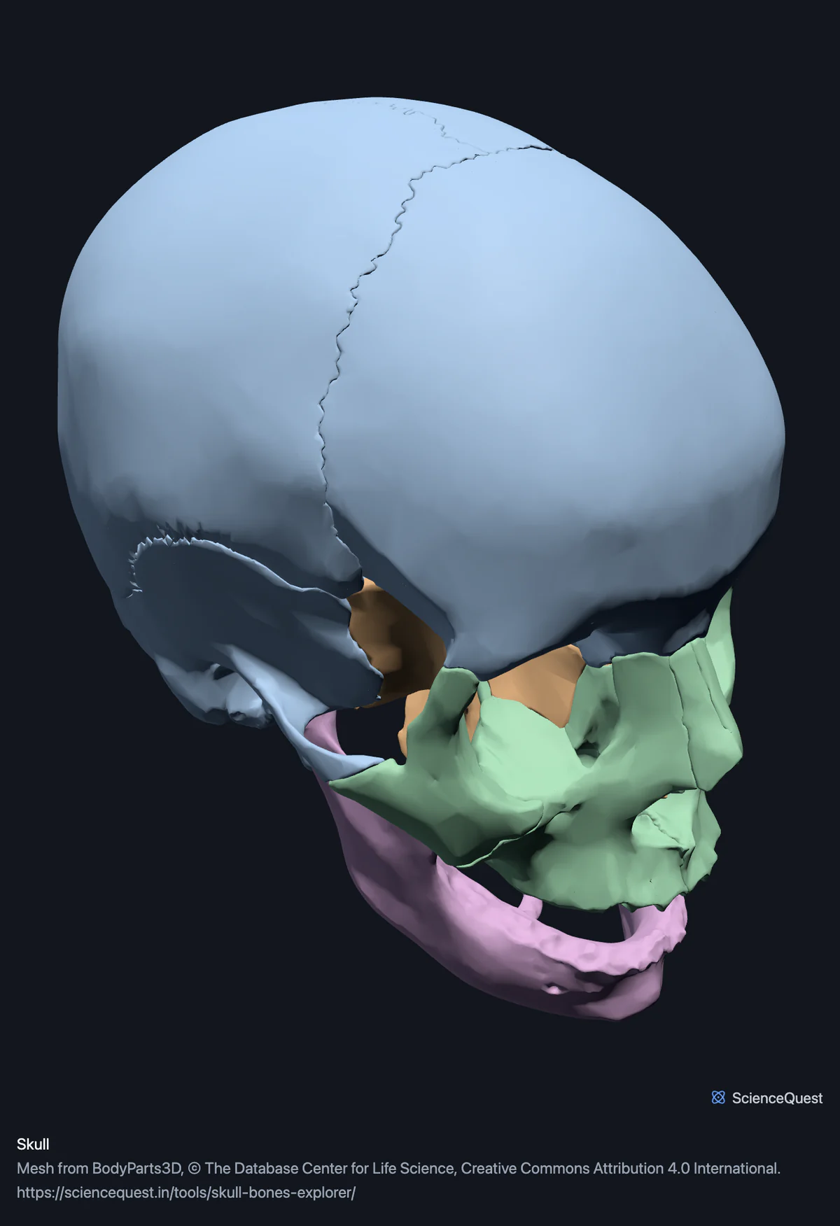
Turn the skull in 3D and take the 22 bones apart one at a time. The sutured vault separates, which is the thing an intact specimen cannot show you.
Visualiser
Skull, 22 bones in 23 meshes
Checking this device for a stored copy.
Start here
- Hide the Cranial vault group Leaves the base and the face, so the sphenoid and the ethmoid are visible for the first time. On an intact skull both are almost entirely hidden.
- Select the sphenoid bone and switch on Isolate selection It spans the whole base and reaches into both orbits, which is why it cannot be learned from any single view of a real specimen.
- Turn to Base, then switch on Cut plane Shows the floor of the cranial cavity and how the palatine bones and the maxillae form the hard palate below it.
Bone only. No teeth, no auditory ossicles and no sutural detail beyond where the segmented surfaces meet. The ossicles sit inside the temporal bones and are not in this dataset, which is one reason the bone count here is 22 rather than 28.
- 22 bones
- 249k triangles
- 1250 KB, on request
- 0.00158 mm worst error per axis
- 6 cranial vault
- 3 skull base
- 12 facial bones
- 2 jaw and hyoid
The hyoid bone loads with them for context and is not counted above. 23 meshes load in total, and every one of them is listed, nameable and selectable.
22 bones, within 23 meshes. The standard division is eight cranial and fourteen facial, and it cuts across the bands here, which group by where a bone sits rather than by what it is. The eight cranial bones are the six in the Cranial vault band plus the sphenoid and the ethmoid from Skull base. The fourteen facial bones are the twelve in Facial bones plus the vomer, which is also in Skull base, and the mandible. The twenty-third mesh is the hyoid, a bone of the neck rather than the skull. Counts of 28 add the six auditory ossicles inside the temporal bones, which BodyParts3D does not publish. A newborn’s count is higher again, because several of these fuse during childhood.
Where this comes from
Segmented from imaging of one adult male body, not drawn by an artist, so the shapes are measurements. Two consequences worth knowing: it is one person’s anatomy rather than an average, and faint stepping on some surfaces is the resolution of the original scan rather than a rendering fault.
Meshes from BodyParts3D, © The Database Center for Life Science, under Creative Commons Attribution 4.0 International. Names and identifiers are the dataset’s own: Foundational Model of Anatomy identifiers and English labels, as its parts list gives them. Converted meshes are shared under the same licence.
Changes made: Meshes were converted from binary STL to an indexed mesh with 16-bit quantised vertex positions, and surface normals were recomputed from the geometry. No vertex was moved other than by that quantisation, whose worst-case error on any one coordinate is reported on the page.
Latin terms for 22 of the 23 structures come from English Wikipedia anatomy infoboxes, under Creative Commons Attribution-ShareAlike 4.0, joined by Wikidata property P1402, Foundational Model of Anatomy identifier. Only a term a source states for that structure is used, never one constructed from a family name, which is why the rest carry no Latin rather than a guess.
All 23 structures
Every structure the model contains, group by group. Of these, 22 are bones of this region. Each name opens the viewer with that structure selected, so a link can point at one structure rather than at the whole region.
Cranial vault 6
- frontal bone os frontale
- right parietal bone os parietale
- left parietal bone os parietale
- right temporal bone os temporale
- left temporal bone os temporale
- occipital bone os occipitale
Skull base 3
- sphenoid bone os sphenoidale
- ethmoid os ethmoidale
- vomer vomer
Facial bones 12
- right maxilla maxilla
- left maxilla maxilla
- right zygomatic bone os zygomaticum, zygoma
- left zygomatic bone os zygomaticum, zygoma
- right nasal bone os nasale
- left nasal bone os nasale
- right lacrimal bone os lacrimale
- left lacrimal bone os lacrimale
- right palatine bone os palatinum
- left palatine bone os palatinum
- right inferior nasal concha concha nasi inferior, concha nasalis inferior
- left inferior nasal concha concha nasi inferior, concha nasalis inferior
Jaw and hyoid 2
- mandible
- hyoid bone os hyoideum
Citing this tool
Last updated . Add the date you accessed it as well, which a citation of a page that can change asks for. If a specific view matters, cite the page address as it stands in your browser instead: it records what is selected and shown.
Eight cranial, fourteen facial
That is the standard division of the 22, and it is worth writing out because it does not match the colours in the viewer:
- Cranial, eight
- Frontal, two parietals, two temporals, occipital, sphenoid and ethmoid. The bones that enclose the brain.
- Facial, fourteen
- Two each of maxilla, zygomatic, nasal, lacrimal, palatine and inferior nasal concha, plus the vomer and the mandible.
The colour bands group by where a bone sits instead, because that is what helps while looking at it. The sphenoid and the ethmoid are counted as cranial but lie under the vault rather than around it, and the vomer is counted as facial while sitting on the midline of the base, so all three are banded with the base here. The count is a convention. The positions are not.
The hyoid is here and is not a skull bone
It is included because it is the only bone in the body that articulates with nothing at all. It hangs in the neck from muscles and ligaments, below the mandible, touching no other bone. Select it and turn the model to see the gap.
Common questions
How many bones are in the skull?
Twenty-two, and the count is a convention rather than a measurement. The usual division is eight cranial bones enclosing the brain and fourteen facial bones, and this model carries all of them separately. The hyoid is included as a twenty-third part but is not counted as a skull bone: it sits in the neck. Counts of 28 add the six auditory ossicles inside the temporal bones, which are not in this dataset, and a newborn’s count is higher still because several bones here fuse during childhood.
Why is the sphenoid so hard to picture?
Because it meets every other cranial bone and several of the facial ones, twelve bones in all, and no single view shows it. It sits across the base like a butterfly, forms part of each orbit, and most of it is hidden inside the head. Isolating it is the whole reason a model like this beats a photograph of an intact skull: on a real specimen the sphenoid is visible only as fragments, in the orbits, at the side of the skull behind each eye and in the base, and its shape has to be imagined from them.
Why does the skull come apart here when a real skull is fused?
Because the dataset segments each bone as its own surface, and the sutures are where those surfaces meet. On an adult specimen the vault behaves as one piece, so the parietals and the frontal can only be learned as regions of a single shell. Hiding the frontal bone and looking at what is left is a view no dry skull offers, and it makes the suture lines obvious in a way tracing them on an intact cranium does not.
Which bone articulates with nothing?
The hyoid, which is why it is in this model despite not being a skull bone. Every other bone in the body meets at least one other at a joint. The hyoid is suspended in the neck by muscles and ligaments alone, sitting below the mandible, and that isolation is what makes it the bone examined after a suspected strangulation. Select it and turn the model: there is a clear gap between it and everything else.