Cervical Spine and Skull Base Explorer
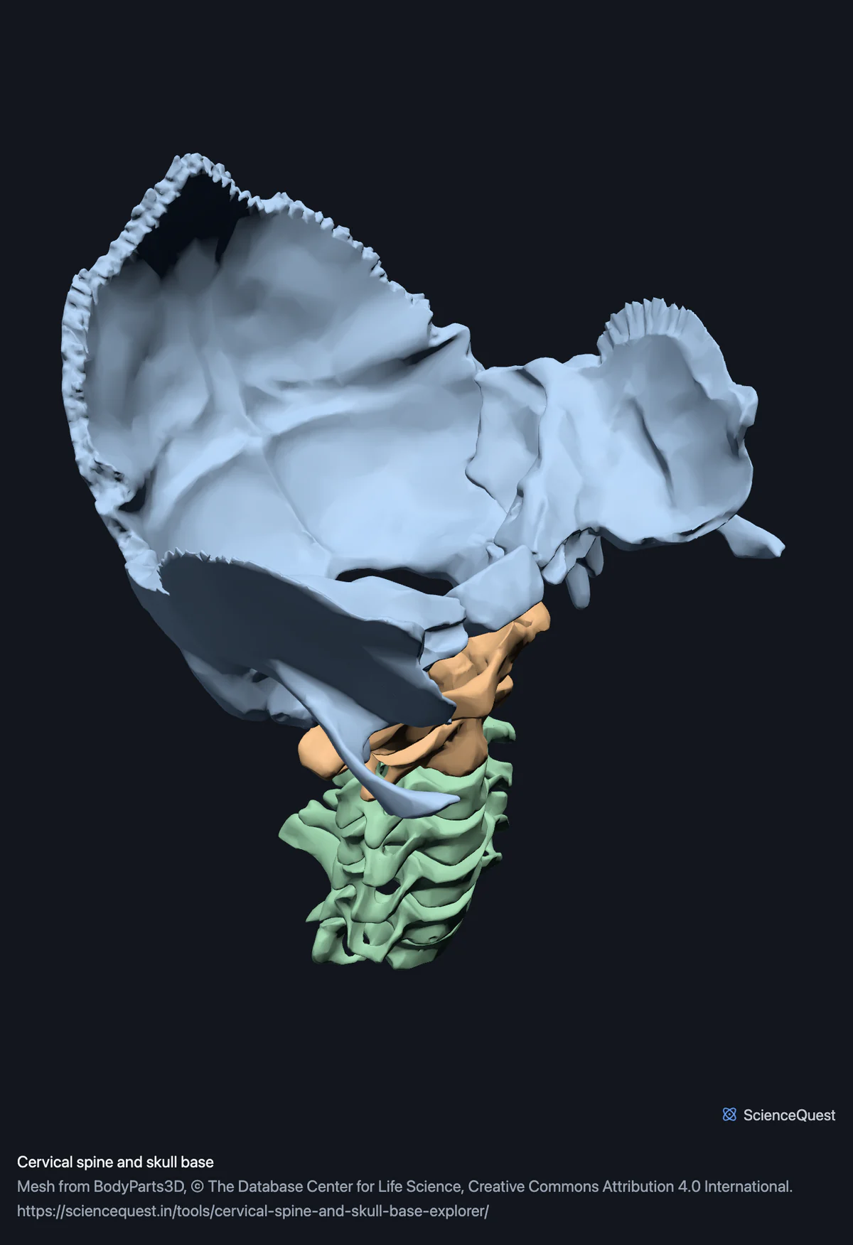
Turn the seven neck vertebrae in 3D with the skull base above them, so the joint that nods the head and the one that rotates it are both visible.
Visualiser
Cervical spine and skull base, 7 bones in 10 meshes
Checking this device for a stored copy.
Start here
- Select the atlas, then the axis One is a ring with no body at all. The other has a peg standing up through that ring. Neither looks like a normal vertebra.
- Hide the Skull base group Leaves the column alone, so the long spinous process of the seventh is unobstructed.
- Turn to Back and compare the third with the seventh The upper processes are short and often split in two. The seventh is the long one you can feel through the skin.
Bone only. No intervertebral discs, which is about a quarter of the height of a real cervical column, so these vertebrae sit closer together than yours do. No ligaments, no spinal cord and no vertebral arteries, and the transverse foramina they pass through are visible but empty.
- 7 bones
- 89k triangles
- 470 KB, on request
- 0.0016 mm worst error per axis
- 3 skull base
- 2 atlas and axis
- 5 cervical vertebrae
The occipital bone, right temporal bone and left temporal bone load with them for context and are not counted above. 10 meshes load in total, and every one of them is listed, nameable and selectable.
Seven cervical vertebrae, within 10 meshes. The occipital bone and both temporal bones load with them and are skull bones rather than vertebrae: they are here because the joint that nods the head is the occipital bone on the atlas, and the joint that rotates it is the atlas on the axis.
Where this comes from
Segmented from imaging of one adult male body, not drawn by an artist, so the shapes are measurements. Two consequences worth knowing: it is one person’s anatomy rather than an average, and faint stepping on some surfaces is the resolution of the original scan rather than a rendering fault.
Meshes from BodyParts3D, © The Database Center for Life Science, under Creative Commons Attribution 4.0 International. Names and identifiers are the dataset’s own: Foundational Model of Anatomy identifiers and English labels, as its parts list gives them. Converted meshes are shared under the same licence.
Changes made: Meshes were converted from binary STL to an indexed mesh with 16-bit quantised vertex positions, and surface normals were recomputed from the geometry. No vertex was moved other than by that quantisation, whose worst-case error on any one coordinate is reported on the page.
Latin terms for 5 of the 10 structures come from English Wikipedia anatomy infoboxes, under Creative Commons Attribution-ShareAlike 4.0, joined by Wikidata property P1402, Foundational Model of Anatomy identifier. Only a term a source states for that structure is used, never one constructed from a family name, which is why the rest carry no Latin rather than a guess.
All 10 structures
Every structure the model contains, group by group. Of these, 7 are bones of this region. Each name opens the viewer with that structure selected, so a link can point at one structure rather than at the whole region.
Skull base 3
- occipital bone os occipitale
- right temporal bone os temporale
- left temporal bone os temporale
Citing this tool
Last updated . Add the date you accessed it as well, which a citation of a page that can change asks for. If a specific view matters, cite the page address as it stands in your browser instead: it records what is selected and shown.
Two joints, two movements
- Atlanto-occipital
- The atlas against the occipital bone. Nodding. The atlas is a ring with no vertebral body, so it carries the skull on two concave facets that cradle the occipital condyles instead.
- Atlanto-axial
- The atlas pivoting on the dens, the peg rising from the axis below it. Rotation. This is the joint that shakes the head.
The remaining five are conventional vertebrae that flex and extend a little each. Nearly half the range of your neck comes from the two at the top.
Why the skull base is here
The nodding joint is between the atlas and the occipital bone, so a neck that stopped below the skull could not show it. The paired temporal bones also fix which side is which, which no two vertebrae could: they all sit on the midline.
Common questions
What makes the atlas and the axis different from the other five?
They are built for two different movements and neither looks much like a normal vertebra. The atlas is a ring with no body at all: it carries the skull on two concave facets that cradle the occipital condyles, and the joint it makes with the occipital bone is what lets you nod. The axis below it has a peg, the dens, standing up through that ring, and the atlas pivots around it, which is what lets you shake your head. Isolate each in turn and the division of labour is obvious, which is the thing a diagram struggles to convey.
Why does this model include part of the skull?
Two reasons, and both are worth stating. The anatomy is the first: the joint that nods the head is between the atlas and the occipital bone, so a neck that stops below the skull cannot show it, and the mastoid processes of the temporal bones are where much of the neck attaches. The second is technical. Every vertebra sits on the midline, so no pair of them can establish left from right, and a view button marked Left would have been a guess. The paired temporal bones settle it, and a skull viewed from either side is symmetrical enough that nothing else would have caught the error.
Why is the seventh cervical vertebra the one you can feel?
Because its spinous process is much longer than the ones above it, which is why it is also called the vertebra prominens: it is the bump at the base of the neck when you bend your head forward. Turn to the back view and compare it with the third or fourth, where the processes are short and often split in two. The length is what makes it the reference point clinicians count ribs and vertebrae from.
Are the intervertebral discs included?
No. BodyParts3D does publish discs as separate structures, and the full skeleton model carries them, but this region is bone only, so the vertebrae here stand apart with nothing between them. That matters for interpreting what you see: roughly a quarter of the height of a real cervical column is disc, and the gaps you can see between these bones are where that tissue belongs rather than empty space.