Occipital bone
The occipital bone is the single bone that forms the back of the skull and the back of the cranial floor. StatPearls calls it the most posterior cranial bone. (OpenStax A&P 2e, 7.2; StatPearls, Occipital Bone, Artery, Vein, and Nerve)
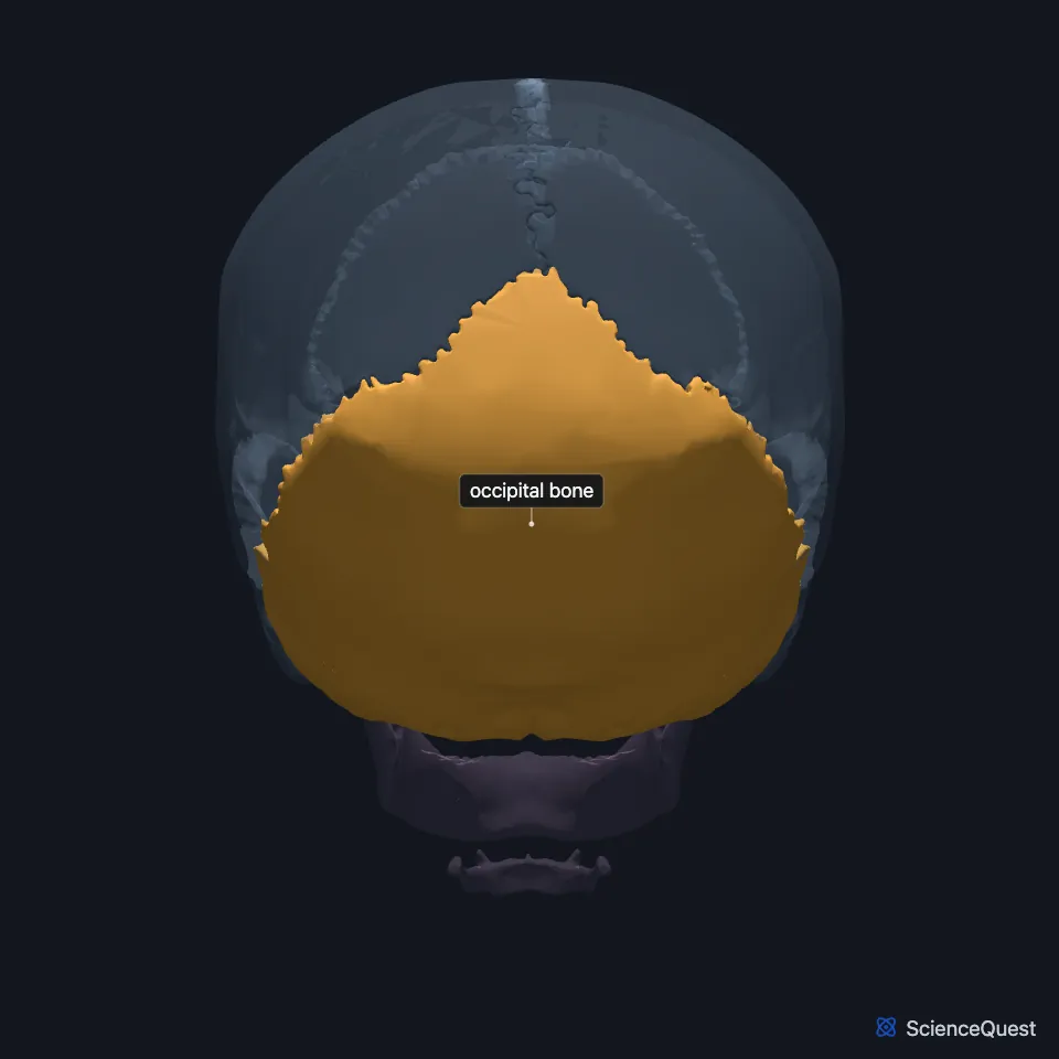
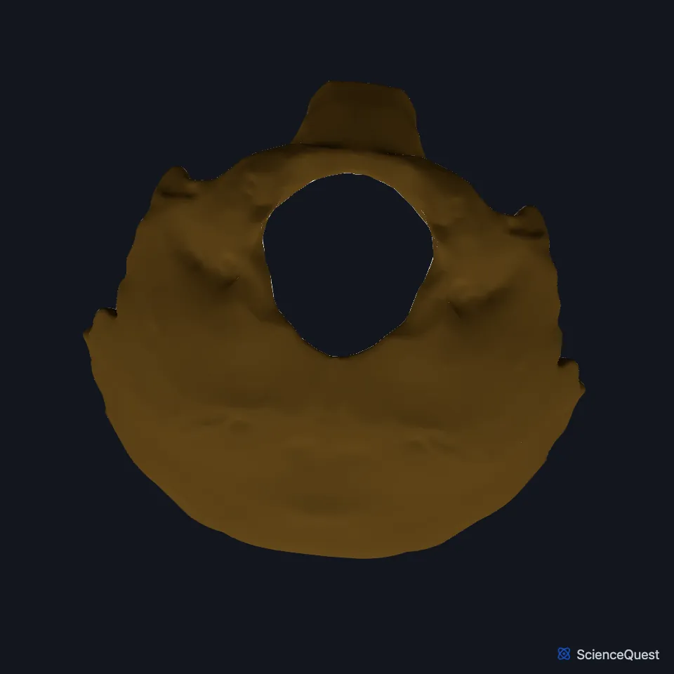
Where is the occipital bone?
It is the rearmost bone of the skull and curves under onto the skull base around the foramen magnum. Its inner surface forms the base of the posterior cranial fossa, which holds the cerebellum. (OpenStax A&P 2e, 7.2; StatPearls, Skull and Occipital Bone, Artery, Vein, and Nerve)
Its outer surface has a midline prominence, the external occipital protuberance, with a superior nuchal line running out to each side. Neck muscles such as the semispinalis capitis attach below that line. (OpenStax A&P 2e, 7.2; StatPearls, Occipital Bone, Artery, Vein, and Nerve)
What are the parts of the occipital bone?
StatPearls divides it into three parts. The squamous part is the largest and most posterior, the condylar parts flank the foramen magnum, and the basilar part lies in front of the foramen and joins the sphenoid. (StatPearls, Occipital Bone, Artery, Vein, and Nerve)
The hypoglossal canal lies at the base of each occipital condyle, beside the foramen magnum, and carries the hypoglossal nerve to the tongue. (OpenStax A&P 2e, 7.2; StatPearls, Occipital Bone, Artery, Vein, and Nerve)
The bone forms from five elements: four of cartilage, the basilar, two condylar and the squamous, and one of membrane, the interparietal. They unite around the foramen magnum. (StatPearls, Foramen Magnum)
What does the occipital bone do?
It protects the cerebellum and the occipital lobes of the cerebrum. On its inner surface, two upper fossae hold the occipital lobes and two lower fossae hold the cerebellar hemispheres. (StatPearls, Occipital Bone, Artery, Vein, and Nerve)
The occipital condyles, one on each side of the foramen magnum, are the skull’s joint surfaces for the superior facets of the atlas, the first cervical vertebra. (OpenStax A&P 2e, 7.2; StatPearls, Occipital Bone, Artery, Vein, and Nerve)
The foramen magnum, the largest opening of the skull, is the passage through which the brain connects to the spinal cord. (StatPearls, Foramen Magnum)
What attaches to the occipital bone?
Neck muscles attach along the nuchal lines. The semispinalis capitis attaches on the lower medial side of the superior nuchal line, and the obliquus capitis superior further out, on the bone between the superior and inferior nuchal lines. The trapezius and the nuchal ligament attach at the inion, the top of the external occipital protuberance, and along the external occipital crest, the midline ridge that runs down to the foramen magnum. (StatPearls, Occipital Bone, Artery, Vein, and Nerve and Suboccipital Muscles)
The rectus capitis posterior major arises from the spinous process of the axis, and the minor from the posterior tubercle of the atlas. Both insert on the inferior nuchal line. (StatPearls, Suboccipital Muscles)
It meets the parietal bones at the lambdoid suture and the mastoid parts of the temporal bones at the occipitomastoid sutures. In front it joins the sphenoid, and below it joins the atlas. (StatPearls, Occipital Bone, Artery, Vein, and Nerve)
Where the sources differ
OpenStax describes the foramen magnum as the passage for the spinal cord as it leaves the skull. StatPearls lists the medulla oblongata among the structures that pass through it, with the accessory nerves, the vertebral and spinal arteries, the alar ligaments and the tectorial membrane. (OpenStax A&P 2e, 7.2; StatPearls, Occipital Bone, Artery, Vein, and Nerve)
| Feature | Detail and source |
|---|---|
| Parts | Squamous, condylar and basilar (StatPearls, Occipital Bone, Artery, Vein, and Nerve) |
| Articulates with | Parietal bones, temporal bones, sphenoid, atlas (StatPearls, Occipital Bone, Artery, Vein, and Nerve) |
| Openings | Foramen magnum, hypoglossal canals; the jugular foramen with the temporal bone (OpenStax A&P 2e, 7.2; StatPearls, Occipital Bone, Artery, Vein, and Nerve) |
Touches in this model
Measured on the BodyParts3D meshes: surfaces within 2 mm of the occipital bone, nearest first, 12 of 42.
- Left parietal bone in contact
- Left temporal bone in contact
- Right parietal bone in contact
- Right temporal bone in contact
- Sphenoid bone in contact
- Aponeurosis of epicranius in contact
- Descending part of left trapezius in contact
- Descending part of right trapezius in contact
- Left longus capitis in contact
- Left middle temporal gyrus in contact
- Left obliquus capitis superior in contact
- Left occipitalis in contact
Sources
- OpenStax Anatomy and Physiology 2e, section 7.2, The Skull. CC BY-NC-SA 4.0. Read 2026-10-25.
- StatPearls, Anatomy, Head and Neck, Occipital Bone, Artery, Vein, and Nerve. Alex M. Germann; Varun Kashyap. Last update July 24, 2023. CC BY-NC-ND 4.0. Read 2026-10-25.
- StatPearls, Anatomy, Head and Neck, Skull. Bradley W. Anderson; Michael W. Kortz; Asa C. Black; Khalid A. Al Kharazi. Last update November 9, 2023. CC BY-NC-ND 4.0. Read 2026-10-25.
- StatPearls, Anatomy, Head and Neck: Foramen Magnum. Jennifer Ficke; Felix Jozsa. Last update April 11, 2026. CC BY-NC-ND 4.0. Read 2026-10-25.
- StatPearls, Anatomy, Head and Neck, Suboccipital Muscles. Tom George; Prasanna Tadi. Last update January 1, 2023. CC BY-NC-ND 4.0. Read 2026-10-25.
The values are facts and free to use; this page’s selection and presentation are © 2026 ScienceQuest.