Sphenoid bone
The sphenoid is a single, complex bone at the center of the skull, forming part of its base and sides. Because it joins so many bones, it helps hold the skull rigid. (OpenStax A&P 2e, 7.2; StatPearls, Sphenoid Bone; Gray 1918, 5. The Skull)
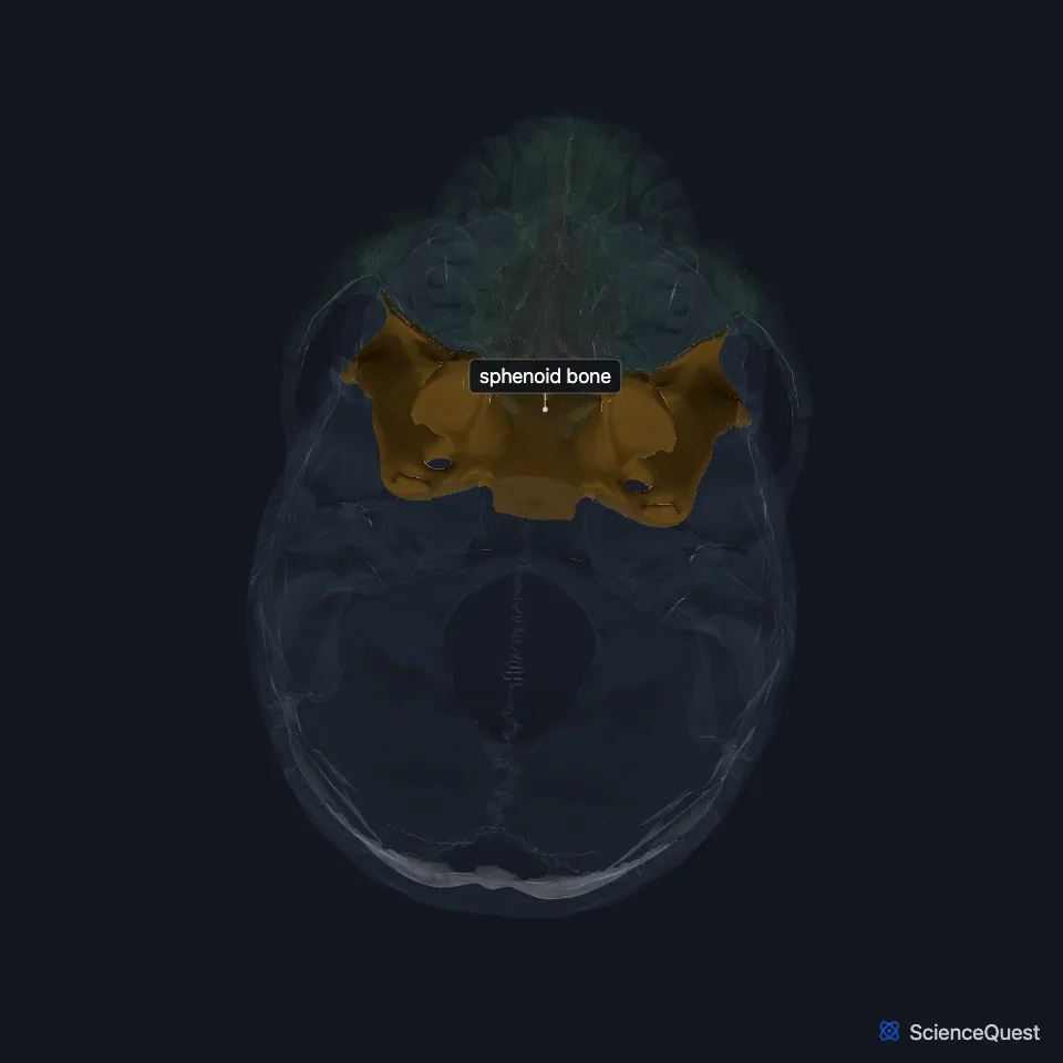
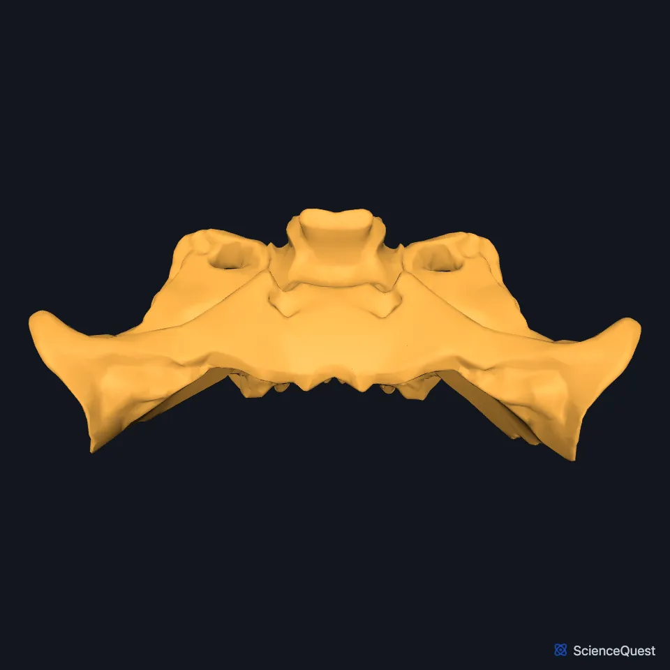
Where is the sphenoid bone?
It lies in the midline of the cranial cavity, behind the frontal bone and in front of the occipital bone. It forms much of the central skull base and reaches out to the sides of the skull. (StatPearls, Sphenoid Bone; OpenStax A&P 2e, 7.2)
On the side of the skull, the greater wing has a temporal surface where the temporalis muscle attaches. In the orbit, the greater wing and the zygomatic bone form the lateral wall. (OpenStax A&P 2e, 7.2; StatPearls, Sphenoid Bone and Orbit Bones)
What are the parts of the sphenoid?
It has a central body, two greater wings, two lesser wings and the downward pterygoid processes. StatPearls compares its shape to a butterfly or a bat. (StatPearls, Sphenoid Bone)
On top of the body, the sella turcica has a central hollow that holds the pituitary gland. (OpenStax A&P 2e, 7.2; StatPearls, Sphenoid Bone)
Below, each pterygoid process points downward as two plates, the medial and lateral pterygoid plates. The lateral pterygoid muscle attaches to the outer face of the lateral plate. (OpenStax A&P 2e, 7.2; StatPearls, Sphenoid Bone)
What does the sphenoid do?
Inside the skull, the back edges of the lesser wings mark the rear limit of the anterior cranial fossa. Behind them, the sphenoid and the two temporal bones form the middle cranial fossa. (OpenStax A&P 2e, 7.2; Gray 1918, 5d. The Interior of the Skull; StatPearls, Skull)
Its openings carry nerves and vessels into and out of the skull: the optic canal, the superior orbital fissure, the foramen rotundum, the foramen ovale and the foramen spinosum, and StatPearls also counts the foramen lacerum, which lies between the sphenoid, occipital and temporal bones. The optic canal passes the optic nerve into the orbit. (OpenStax A&P 2e, 7.2; StatPearls, Sphenoid Bone)
StatPearls credits its many joints with other bones for the rigidity of the skull, and names it as an attachment site for many of the muscles of mastication. (StatPearls, Sphenoid Bone)
Which bones and muscles meet the sphenoid?
It articulates with the frontal, parietal, ethmoid, zygomatic, temporal, occipital, palatine and vomer bones. (StatPearls, Sphenoid Bone)
The temporalis attaches to the temporal surface of the greater wing. The upper fibers of the lateral pterygoid attach to the infratemporal surface of the greater wing, and the lateral pterygoid and medial pterygoid attach to the two sides of the lateral pterygoid plate. (StatPearls, Sphenoid Bone)
At the pterion, the greater wing meets the frontal, parietal and temporal bones. (OpenStax A&P 2e, 7.2; StatPearls, Skull; Gray 1918, 5d. The Interior of the Skull)
Where the sources differ
OpenStax describes the sphenoid sinus as a single midline sinus in the body of the sphenoid, the most posterior of the paranasal sinuses. StatPearls refers to sphenoidal sinuses, in the plural, in the body of the bone. (OpenStax A&P 2e, 7.2; StatPearls, Sphenoid Bone)
| Feature | Detail and source |
|---|---|
| Parts | Body, greater wings, lesser wings and pterygoid processes (StatPearls, Sphenoid Bone) |
| Articulates with | Frontal, parietal, ethmoid, zygomatic, temporal, occipital, palatine and vomer bones (StatPearls, Sphenoid Bone) |
| Openings | Optic canal, superior orbital fissure, foramen rotundum, foramen ovale, foramen spinosum; StatPearls also counts the foramen lacerum (OpenStax A&P 2e, 7.2; StatPearls, Sphenoid Bone) |
Touches in this model
Measured on the BodyParts3D meshes: surfaces within 2 mm of the sphenoid bone, nearest first, 12 of 38.
- Ethmoid in contact
- Frontal bone in contact
- Left maxilla in contact
- Left palatine bone in contact
- Left parietal bone in contact
- Left temporal bone in contact
- Left zygomatic bone in contact
- Mandible in contact
- Occipital bone in contact
- Right palatine bone in contact
- Right parietal bone in contact
- Right temporal bone in contact
Sources
- OpenStax Anatomy and Physiology 2e, section 7.2, The Skull. CC BY-NC-SA 4.0. Read 2026-10-25.
- StatPearls, Anatomy, Sphenoid Bone. Radia T. Jamil; Avery L. Callahan. Last update July 24, 2023. CC BY-NC-ND 4.0. Read 2026-10-25.
- Gray’s Anatomy of the Human Body, 20th US edition (1918), II. Osteology, 5. The Skull, opening list of the cranial and facial bones. Henry Gray. Public domain. Read 2026-10-25.
- StatPearls, Anatomy, Head and Neck, Orbit Bones. Caleb L. Shumway; Mahsaw Motlagh; Matthew Wade. Last update July 24, 2023. CC BY-NC-ND 4.0. Read 2026-10-25.
- Gray’s Anatomy of the Human Body, 20th US edition (1918), II. Osteology, 5d. The Interior of the Skull, Upper Surface of the Base of the Skull, and the table of craniometric points (1918 terms: small wings and great wing for lesser and greater wings). Henry Gray. Public domain. Read 2026-10-25.
- StatPearls, Anatomy, Head and Neck, Skull. Bradley W. Anderson; Michael W. Kortz; Asa C. Black; Khalid A. Al Kharazi. Last update November 9, 2023. CC BY-NC-ND 4.0. Read 2026-10-25.
The values are facts and free to use; this page’s selection and presentation are © 2026 ScienceQuest.