Maxilla
The maxillae are a pair of bones that together form the upper jaw, much of the hard palate, the medial floor of the orbit and the lateral base of the nose. Each holds the maxillary sinus, the largest paranasal sinus. (OpenStax A&P 2e, 7.2; StatPearls, Maxilla)
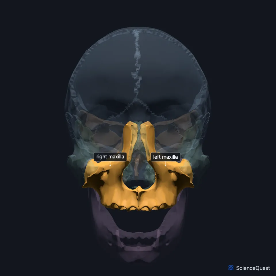
Where is the maxilla?
Each maxilla sits in the middle of the face, below the orbit. It meets the frontal bone above, the zygomatic bone to the side and the palatine bone behind. (OpenStax A&P 2e, 7.2; StatPearls, Maxilla)
At the front, it forms the lower and side margins of the nasal opening, the pyriform aperture, and meets the nasal bones along its frontal process. (StatPearls, Maxilla)
What are the parts of the maxilla?
Each maxilla has a pyramidal body and four processes: alveolar, frontal, zygomatic and palatine. (StatPearls, Maxilla)
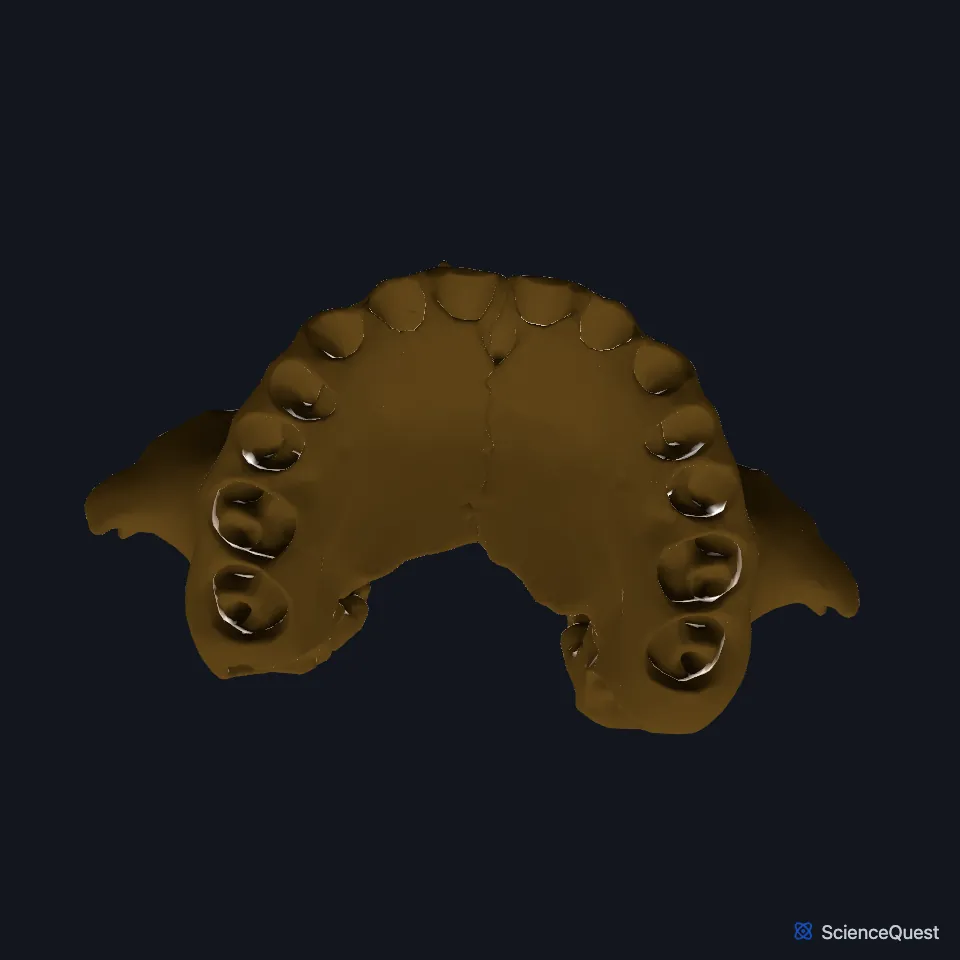
The alveolar process, a horseshoe-shaped ridge below the hard palate, anchors the upper teeth. (OpenStax A&P 2e, 7.2; StatPearls, Maxilla)
The maxillary sinus fills the hollow body and has an adult volume of about 15 mL. Its roof forms part of the orbital floor and carries the infraorbital canal. (StatPearls, Maxilla)
What does the maxilla do?
It anchors the upper teeth and forms most of the hard palate, the bony plate that separates the mouth from the nasal cavity. Chewing forces pass through its frontal and zygomatic processes to the cranium. (OpenStax A&P 2e, 7.2; StatPearls, Maxilla)
The infraorbital foramen, just below the orbit, is where a sensory nerve to the nose, upper lip and front of the cheek leaves the bone. The maxillary nerve, the second division of the trigeminal nerve, supplies the maxilla. (OpenStax A&P 2e, 7.2; StatPearls, Maxilla)
What joins and attaches to the maxilla?
The right and left maxillae meet in the midline through their palatine processes, at the median maxillary suture. Behind them, the horizontal plates of the palatine bones form the back quarter of the hard palate. (StatPearls, Maxilla; OpenStax A&P 2e, 7.2)
In the orbit, the maxilla forms most of the floor. The palatine bone and the zygomatic bone complete it. (OpenStax A&P 2e, 7.2; StatPearls, Maxilla and Orbit Bones)
The nasalis, levator labii superioris alaeque nasi, levator labii superioris and levator anguli oris arise from the maxilla. Both levator labii superioris muscles run down into the orbicularis oris. The maxillary bundle of the buccinator arises from the cheek side of the alveolar process. (StatPearls, Maxilla and Buccinator Muscle)
The superficial head of the medial pterygoid, a chewing muscle, arises from the maxillary tuberosity. (StatPearls, Mastication Muscles)
How does the maxilla form?
In the embryo, the maxillary prominences develop into the secondary palate, most of the maxilla and the side of the upper lip. Palatal fusion is complete by the 10th week. (StatPearls, Maxilla)
Where the sources differ
The sources give different shares of the hard palate to the palatine processes of the maxillae. OpenStax says they form the anterior three quarters; StatPearls says two thirds. (OpenStax A&P 2e, 7.2; StatPearls, Maxilla)
Of the midface muscles, the StatPearls maxilla article says only the nasalis, levator labii superioris alaeque nasi, levator labii superioris and levator anguli oris arise from the maxilla. The StatPearls buccinator article gives the buccinator a maxillary bundle that arises from the alveolar process of the maxilla. (StatPearls, Maxilla and Buccinator Muscle)
| Feature | Detail and source |
|---|---|
| Parts | Body and four processes: alveolar, frontal, zygomatic, palatine (StatPearls, Maxilla) |
| Articulates with, among others | The other maxilla, frontal bone, ethmoid, zygomatic bone, palatine bone, nasal bones, inferior nasal concha (StatPearls, Maxilla and Ethmoid Bone) |
| Holds | The upper teeth and the maxillary sinus (OpenStax A&P 2e, 7.2; StatPearls, Maxilla) |
Touches in this model
Measured on the BodyParts3D meshes: surfaces within 2 mm of the maxilla, nearest first, 12 of 64.
- Ethmoid in contact
- Frontal bone in contact
- Left lacrimal bone in contact
- Left nasal bone in contact
- Left palatine bone in contact
- Left zygomatic bone in contact
- Right inferior nasal concha in contact
- Right lacrimal bone in contact
- Right nasal bone in contact
- Right palatine bone in contact
- Right zygomatic bone in contact
- Sphenoid bone in contact
Sources
- OpenStax Anatomy and Physiology 2e, section 7.2, The Skull. CC BY-NC-SA 4.0. Read 2026-10-25.
- StatPearls, Anatomy, Head and Neck, Maxilla. Roberto M. Soriano; Melina Brizuela. Last update June 23, 2025. CC BY-NC-ND 4.0. Read 2026-10-25.
- StatPearls, Anatomy, Head and Neck, Orbit Bones. Caleb L. Shumway; Mahsaw Motlagh; Matthew Wade. Last update July 24, 2023. CC BY-NC-ND 4.0. Read 2026-10-25.
- StatPearls, Anatomy, Head and Neck: Buccinator Muscle. Manu Rathee; Prachi Jain. Last update January 30, 2024. CC BY-NC-ND 4.0. Read 2026-10-25.
- StatPearls, Anatomy, Head and Neck, Mastication Muscles. Hajira Basit; Muhammad Ali Tariq; Marco A. Siccardi. Last update June 5, 2023. CC BY-NC-ND 4.0. Read 2026-10-25.
- StatPearls, Anatomy, Head and Neck, Ethmoid Bone. Megan Yu; Shu-Min Wang. Last update July 24, 2023. CC BY-NC-ND 4.0. Read 2026-10-25.
The values are facts and free to use; this page’s selection and presentation are © 2026 ScienceQuest.