Orbicularis oris
The orbicularis oris is the main muscle of the lips. It has no bony or tendinous origin, and many other facial muscles around the mouth attach to it. (OpenStax A&P 2e, 11.3; StatPearls, Orbicularis Oris Muscle)
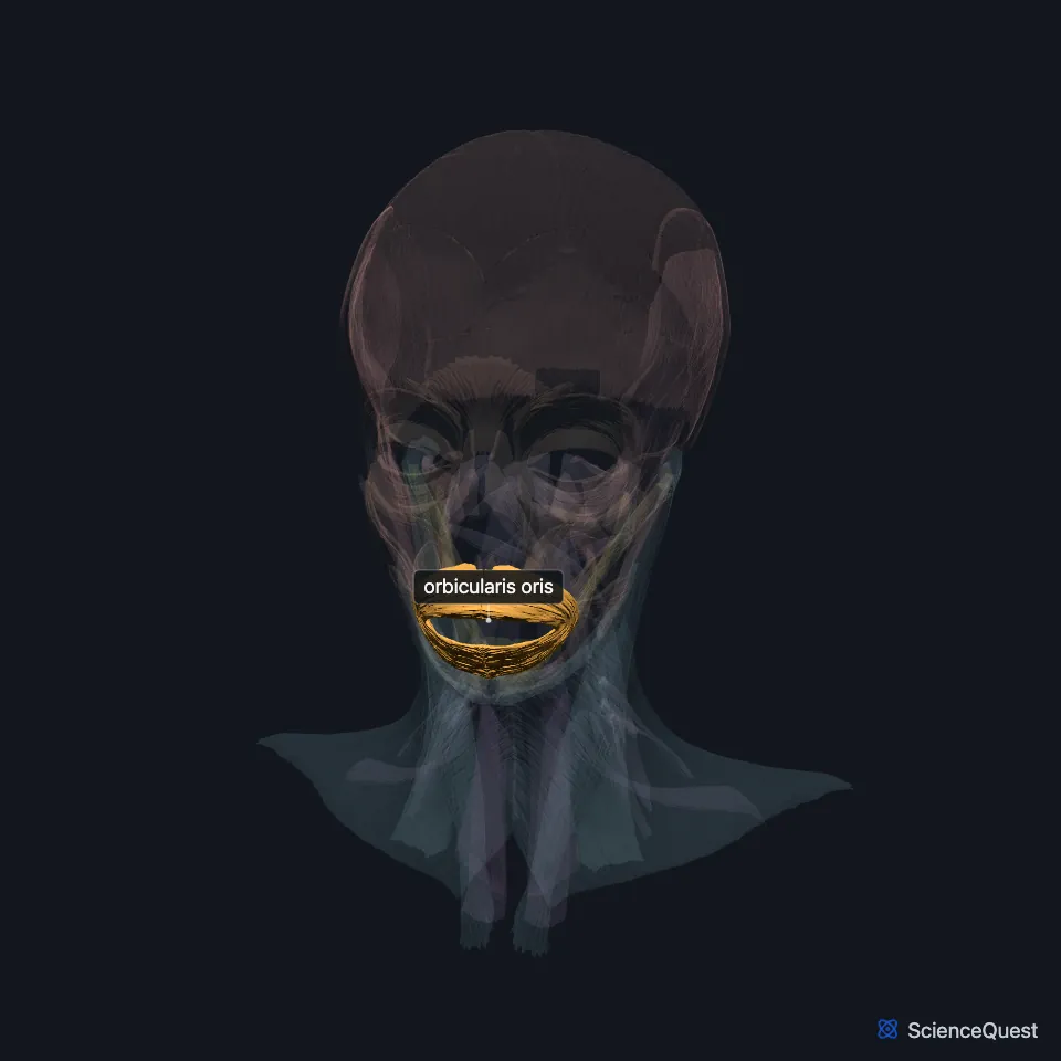
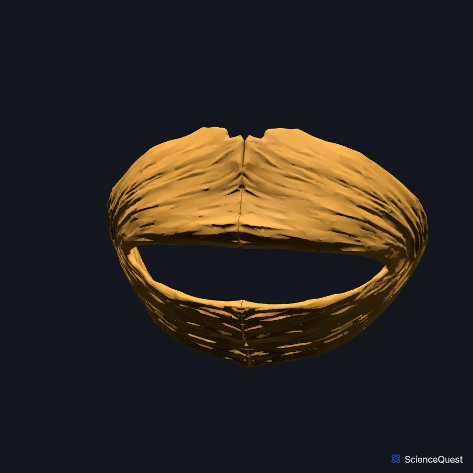
Open it in the Head and Neck Muscles Explorer See it alone in 3D
Where is the orbicularis oris?
It surrounds the mouth and attaches, through a thin superficial musculoaponeurotic layer, to the dermis of the upper and lower lips. (StatPearls, Orbicularis Oris Muscle)
Its deep fibers lie close to the inner lining of the lips and run across the midline from one corner of the mouth to the other. (StatPearls, Orbicularis Oris Muscle)
What are the layers of the orbicularis oris?
It has a deep part and a superficial part. Authors also call them the marginal and peripheral parts, or pars marginalis and pars superficialis. (StatPearls, Orbicularis Oris Muscle)
The deep fibers start at the modiolus on each side, the point beside the corner of the mouth. Their lower border curls on itself and turns the lining outward to form the vermilion of the lip. (StatPearls, Orbicularis Oris Muscle and Buccinator Muscle)
The superficial fibers come from other facial muscles. The nasolabial bundle takes fibers from the depressor anguli oris and ends in the ridges of the philtrum; the nasal bundle, above it, gathers fibers of the zygomaticus major and minor and the levator labii superioris and ends at the base of the nose. (StatPearls, Orbicularis Oris Muscle)
What does the orbicularis oris do?
Its deep fibers act as a constrictor, the sphincter action that closes the mouth and holds food in. Its superficial fibers shape the lips for facial expression and for the precise lip movements of speech. (StatPearls, Orbicularis Oris Muscle)
It puckers the lips, so it is sometimes called the kissing muscle, and players of brass and woodwind instruments rely on it. (StatPearls, Orbicularis Oris Muscle)
With the buccinator and the pharyngeal constrictors it forms the buccinator mechanism, used in swallowing, sucking, whistling, chewing and saying vowels. (StatPearls, Orbicularis Oris Muscle)
What supplies the orbicularis oris, and what joins it?
The buccal and marginal mandibular branches of the facial nerve supply it. Its main blood supply is the facial artery, through its superior and inferior labial branches. (StatPearls, Orbicularis Oris Muscle and Orbicularis Oculi Muscle; FMA, Marginal mandibular branch of facial nerve)
The buccinator’s fibers blend into it, and part of the platysma inserts into it. (StatPearls, Buccinator Muscle and Platysma)
How does the orbicularis oris vary?
Some babies are born without it on one side of the face, which leaves part of that side drooping. (StatPearls, Orbicularis Oris Muscle)
Tapping beside the nose or on the upper lip of an infant raises the corner of the mouth on that side. This orbicularis oris reflex, or snout reflex, disappears later in life. (StatPearls, Orbicularis Oris Muscle)
Where the sources differ
OpenStax calls it a circular muscle. StatPearls says the older view of it as a sphincter ringing the mouth was dropped once research showed its fibers run in different directions, though it still credits the deep fibers with the mouth’s sphincter action. (OpenStax A&P 2e, 11.3; StatPearls, Orbicularis Oris Muscle)
| Feature | Detail and source |
|---|---|
| Origin | No bony or tendinous origin: deep fibers from the modiolus, superficial fibers from other facial muscles (StatPearls, Orbicularis Oris Muscle) |
| Insertion | Dermis of the lips; ridges of the philtrum; base of the nose (StatPearls, Orbicularis Oris Muscle) |
| Action | Moves, closes and puckers the lips (OpenStax A&P 2e, 11.3; StatPearls, Orbicularis Oris Muscle) |
| Nerve | Facial nerve: buccal and marginal mandibular branches (StatPearls, Orbicularis Oris Muscle; FMA, Marginal mandibular branch of facial nerve) |
Touches in this model
Measured on the BodyParts3D meshes: surfaces within 2 mm of the orbicularis oris, nearest first, 12 of 44.
- Left buccinator in contact
- Left depressor anguli oris in contact
- Left depressor labii inferioris in contact
- Left depressor septi nasi in contact
- Left levator anguli oris in contact
- Left levator labii superioris in contact
- Left risorius in contact
- Left zygomaticus major in contact
- Left zygomaticus minor in contact
- Right buccinator in contact
- Right depressor anguli oris in contact
- Right depressor labii inferioris in contact
Sources
- OpenStax Anatomy and Physiology 2e, section 11.3, Axial Muscles of the Head, Neck, and Back. CC BY-NC-SA 4.0. Read 2026-10-25.
- StatPearls, Anatomy, Head and Neck, Orbicularis Oris Muscle. Prachi Jain; Manu Rathee. Last update August 7, 2023. CC BY-NC-ND 4.0. Read 2026-10-25.
- StatPearls, Anatomy, Head and Neck: Buccinator Muscle. Manu Rathee; Prachi Jain. Last update January 30, 2024. CC BY-NC-ND 4.0. Read 2026-10-25.
- Foundational Model of Anatomy, FMA53365, Marginal mandibular branch of facial nerve (read on EBI OLS). FMA release terms, University of Washington. Read 2026-10-25.
- StatPearls, Anatomy, Head and Neck: Orbicularis Oculi Muscle. John Tong; Michael J. Lopez; Adegbenro O. Fakoya; Bhupendra C. Patel. Last update May 25, 2024. CC BY-NC-ND 4.0. Read 2026-10-25.
- StatPearls, Anatomy, Head and Neck, Platysma. Jacob E. Hoerter; Bhupendra C. Patel. Last update August 7, 2023. CC BY-NC-ND 4.0. Read 2026-10-25.
The values are facts and free to use; this page’s selection and presentation are © 2026 ScienceQuest.