Internal Organs Explorer
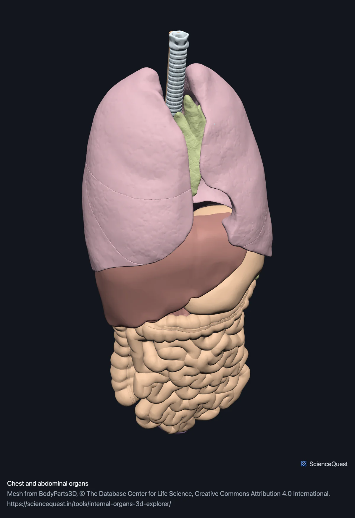
Turn the internal organs in 3D: lungs, airway, the gut from oesophagus to rectum, liver, pancreas, spleen, kidneys and bladder, each one separable.
Visualiser
Chest and abdominal organs, 31 structures
Checking this device for a stored copy.
Start here
- Click Urinary twice, then Front Opening a band frames it, and these bands start open, so the first click folds Urinary and the second takes you to both kidneys and the bladder rather than leaving them small inside a whole torso. The view chips keep whatever is framed.
- Hide Lungs, then Digestive tract, then Liver, gallbladder and pancreas The fastest way to see the kidneys on the back wall of the abdomen. Below the diaphragm what is left is the kidneys, the adrenals sitting on top of them, the ureters running down to the bladder and the spleen against the upper left kidney. The liver band goes too because the liver lies in front of the right kidney and the pancreas in front of the left.
- Select the liver, then turn on Fade the rest Shows how far the liver crosses the midline and how much of the stomach it covers, which is the relationship most diagrams idealise.
Organs, and the three muscle bands of the colon. No heart and no blood vessels: the heart and the great vessels each have a model of their own, and the pancreatic duct in the liver band carries digestive juice, not blood. No mesentery, peritoneum or greater omentum, so the gut floats without the tissue that holds it in place. The dataset publishes none of those as part of these organs.
- 31 structures
- 819k triangles
- 4400 KB, on request
- 0.00504 mm worst error per axis
- 5 lungs
- 2 airway
- 10 digestive tract
- 4 liver, gallbladder and pancreas
- 5 urinary
- 5 spleen, adrenals and thymus
Where this comes from
Segmented from imaging of one adult male body, not drawn by an artist, so the shapes are measurements. Two consequences worth knowing: it is one person’s anatomy rather than an average, and faint stepping on some surfaces is the resolution of the original scan rather than a rendering fault.
Meshes from BodyParts3D, © The Database Center for Life Science, under Creative Commons Attribution 4.0 International. Names and identifiers are the dataset’s own: Foundational Model of Anatomy identifiers and English labels, as its parts list gives them. Converted meshes are shared under the same licence.
Changes made: Meshes were converted from binary STL to an indexed mesh with 16-bit quantised vertex positions, and surface normals were recomputed from the geometry. No vertex was moved other than by that quantisation, whose worst-case error on any one coordinate is reported on the page.
Latin terms for 17 of the 31 structures come from English Wikipedia anatomy infoboxes, under Creative Commons Attribution-ShareAlike 4.0, joined by Wikidata property P1402, Foundational Model of Anatomy identifier. Only a term a source states for that structure is used, never one constructed from a family name, which is why the rest carry no Latin rather than a guess.
All 31 structures
Every structure the model contains, group by group. Each name opens the viewer with that structure selected, so a link can point at one structure rather than at the whole region.
Lungs 5
- upper lobe of right lung organ
- middle lobe of lung organ
- lower lobe of right lung organ
- upper lobe of left lung organ
- lower lobe of left lung organ
Digestive tract 10
- esophagus oesophagus organ
- stomach gaster, ventriculus organ
- duodenum duodenum organ
- jejunum jejunum organ
- ileum ileum organ
- colon organ
- rectum rectum organ
- free taenia muscle
- mesocolic taenia muscle
- omental taenia muscle
Liver, gallbladder and pancreas 4
- liver hepar organ
- gallbladder vesica biliaris, vesica fellea organ
- pancreas organ
- pancreatic duct ductus pancreaticus organ
Urinary 5
- right kidney organ
- left kidney organ
- right ureter ureter organ
- left ureter ureter organ
- urinary bladder vesica urinaria organ
Spleen, adrenals and thymus 5
- spleen splen, lien organ
- right adrenal gland glandula suprarenalis organ
- left adrenal gland glandula suprarenalis organ
- right lobe of thymus organ
- left lobe of thymus organ
Citing this tool
Last updated . Add the date you accessed it as well, which a citation of a page that can change asks for. If a specific view matters, cite the page address as it stands in your browser instead: it records what is selected and shown.
Why this is not a diagram of organs
Every mesh here was segmented from imaging of one adult body, in one shared coordinate frame. So the positions are measurements rather than layout decisions: how far the right lobe of the liver descends over the right kidney, how the left kidney sits higher than the right, how the stomach tucks under the diaphragm and to the left of the midline. A printed illustration is drawn to be legible, which means it separates organs that in life are pressed against each other.
The caveat matters more here than for bone. Abdominal organs move: with posture, with each breath, and with how recently the person ate. A skeleton is more or less where you left it, whereas this is one person’s abdomen at one instant.
The organs had to be named one at a time
The other regions here are generated from the dataset’s own part-of hierarchy, which is what stops a part being dropped by hand. That does not work for organs, and the reason is worth knowing if you use this data yourself: the hierarchy records what a body region contains, not what it is made of.
Taking everything under the respiratory system brings 37 meshes of the mouth with it, the teeth, gums, lips, lower jaw and the muscles of the lips and chin, because the mouth is part of the airway. The alimentary system brings those same 37 plus the masseter and medial pterygoid muscles and the zygomatic bone, because the dataset files them under the cheek. Both are correct statements about anatomy and neither is a list of organs, so each organ below is named explicitly and the meshes beneath it are taken whole.
What the bands mean, and why the colours are not realistic
One hue per system, not per organ. Thirty one separate colours would not stay distinguishable, and realism would lose the information that matters: a real liver and a real spleen are nearly the same dark red, so drawing them honestly would erase the boundary between them, which is the boundary someone examining the upper left abdomen is looking for.
One band is not what its name suggests. The three taeniae coli sit with the digestive tract, and they are tagged as muscle rather than as organs, because that is what they are: the longitudinal smooth muscle bands that gather the colon into its pouches. They are why this model reports three muscle structures alongside its twenty eight organs.
Common questions
Why is the heart not in here?
Because it has its own model, and including it would cost you more than it gave. The wall of the heart is one mesh of 331,592 triangles, which is more than two thirds of the entire heart model and would be close to a third of this download, for one organ that the dedicated heart model already shows far better with its valves, papillary muscles and coronary vessels separable. So this region is everything around the heart, and the heart is one click away.
Which organs are actually here?
Thirty one structures. Five lung lobes and the trachea and bronchial tree. The digestive tract as separate meshes for oesophagus, stomach, duodenum, jejunum, ileum, colon and rectum, along with the three taeniae coli, which are the longitudinal muscle bands of the colon. The liver, gallbladder, pancreas and pancreatic duct. Both kidneys, both ureters and the bladder. The spleen, both adrenal glands and both lobes of the thymus. What is absent is the appendix, which the dataset publishes but this model does not load, and anything the dataset does not publish as its own mesh: there is no mesentery, no peritoneum and no greater omentum, so the gut floats without the tissue that in life holds it in place.
Why are the organs not the colours of real organs?
Because one hue per system is more useful than realism here, and realism would actively lose information. A real liver and a real spleen are nearly the same dark red, so drawing them truthfully would erase the boundary between them, which is exactly the boundary someone looking at the upper left abdomen is trying to find. The colours are loosely conventional, one per system, and the point of them is that the lungs read as one thing and the gut as another.
Is this where the organs really sit?
Yes, and that is the main reason to look at it rather than a diagram. Everything here is segmented from imaging of one adult body, in one shared coordinate frame, so the relationships are measured: how far the liver crosses the midline, how high the left kidney sits above the right, how the stomach lies against the diaphragm. A textbook illustration is drawn for clarity and quietly idealises all of that. The caveat is the same one that comes with any single specimen, and it is worth more for organs than for bones, since abdominal organs shift with posture, breathing and how recently the person ate.