Abdomen and Pelvis Explorer
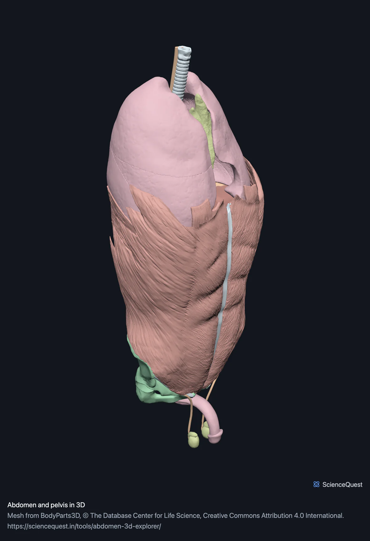
Lumbar spine, pelvis, the abdominal organs, the abdominal wall and the pelvic floor in one 3D scene: 76 structures you can take apart in order.
Visualiser
Lumbar spine and pelvis, 8 bones
Checking this device for a stored copy.
Start here
- Hide the Abdominal wall band Twelve muscles off in one press. The wall is what stops you seeing anything, and hiding it is the closest this model comes to an incision.
- Select a kidney and press Keep only this Then switch the five lumbar vertebrae back on one at a time to find the level it sits at.
- Hide the Lungs band The organ model is the whole trunk, so the lungs come with it. Take them off, and the Airway band after them, and what remains is the abdomen, apart from the thymus and most of the oesophagus, which lie in the chest.
Bone, organs and muscle. No heart, because it is a separate model this page does not load. No nerves, because the dataset’s only nerves anywhere are the optic nerves, with their chiasm and tracts. No lymph nodes or lymphatic vessels, because the catalogue’s single lymphatic entry is the lymphoid system as an undivided whole. The reproductive organs are male, and no female set exists in the catalogue to switch to.
Where this comes from
Segmented from imaging of one adult male body, not drawn by an artist, so the shapes are measurements. Two consequences worth knowing: it is one person’s anatomy rather than an average, and faint stepping on some surfaces is the resolution of the original scan rather than a rendering fault.
Meshes from BodyParts3D, © The Database Center for Life Science, under Creative Commons Attribution 4.0 International. Names and identifiers are the dataset’s own: Foundational Model of Anatomy identifiers and English labels, as its parts list gives them. Converted meshes are shared under the same licence.
Changes made: Meshes were converted from binary STL to an indexed mesh with 16-bit quantised vertex positions, and surface normals were recomputed from the geometry. No vertex was moved other than by that quantisation, whose worst-case error on any one coordinate is reported on the page. On this page it is at most 0.00504 mm, the largest among the 4 models the page can load.
Latin terms for 46 of the 76 structures in these models come from English Wikipedia anatomy infoboxes, under Creative Commons Attribution-ShareAlike 4.0, joined by Wikidata property P1402, Foundational Model of Anatomy identifier. Only a term a source states for that structure is used, never one constructed from a family name, which is why the rest carry no Latin rather than a guess.
Citing this tool
Last updated . Add the date you accessed it as well, which a citation of a page that can change asks for. If a specific view matters, cite the page address as it stands in your browser instead: it records what is selected and shown.
Why the lungs are on a page about the abdomen
Because the dataset segments the chest and abdominal organs as a single model, and this page loads it whole. All five lung lobes arrive with the digestive tract, along with the trachea and the bronchus. They sit in two bands of their own, Lungs and Airway, and come off in two presses, so the page reads as the abdomen the moment you want it to. Loading an asset that has already been checked, rather than re-cutting one into a new binary with a new digest, is what keeps every mesh and every anatomical axis in this scene a verified one. The price is a boundary that follows the segmentation rather than the region, and the page states it instead of hiding structures to make the boundary look tidier than the data is.
Common questions
Why are the lungs in a page about the abdomen?
Because the dataset segments the chest and abdominal organs as one model and this page loads it whole. All five lung lobes arrive with the gut, along with the trachea and the bronchus. They sit in two bands of their own, Lungs and Airway, and come off in two presses, so the page is usable either way. Building it from an asset that is already verified, rather than re-cutting one, is what keeps every mesh, digest and anatomical axis in this scene a checked one, and the price is a boundary that follows the segmentation rather than the region.
What is the best order to take it apart in?
Outside in, which is also the order a dissection goes. Hide the abdominal wall first, because it is the thing stopping you seeing anything, and twelve muscles go in one press. Then the lungs and the airway if you want the abdomen, which leaves only the thymus and most of the oesophagus in the chest. The organs come off band by band after that: digestive tract, then liver and pancreas, which leaves the kidneys against the lumbar spine. Switching the five lumbar vertebrae back on one at a time is how you find the level a kidney sits at.
Are the female reproductive organs available?
No. The BodyParts3D catalogue contains no female reproductive structures at all: no ovary, uterus, uterine tube or vagina appears in it, so there is nothing to load and nothing to switch to. The reproductive layer here is the male set, which the dataset does publish. This is a real gap in the source data rather than a decision made on this site, and it is stated rather than left for a reader to find by searching.
Are the lymph nodes and the nerves here?
Neither, and both absences are in the source rather than in this page. The catalogue’s only lymphatic entry is the lymphoid system as an undivided whole, so there are no nodes and no lymphatic vessels to show. For nerves, the whole dataset publishes the optic nerves, their tracts and the chiasm and nothing else, so there is no lumbar plexus and no autonomic supply. Every model here shows what the source publishes, which means an absence is a fact about the segmentation.