Lower Limb Muscles Explorer
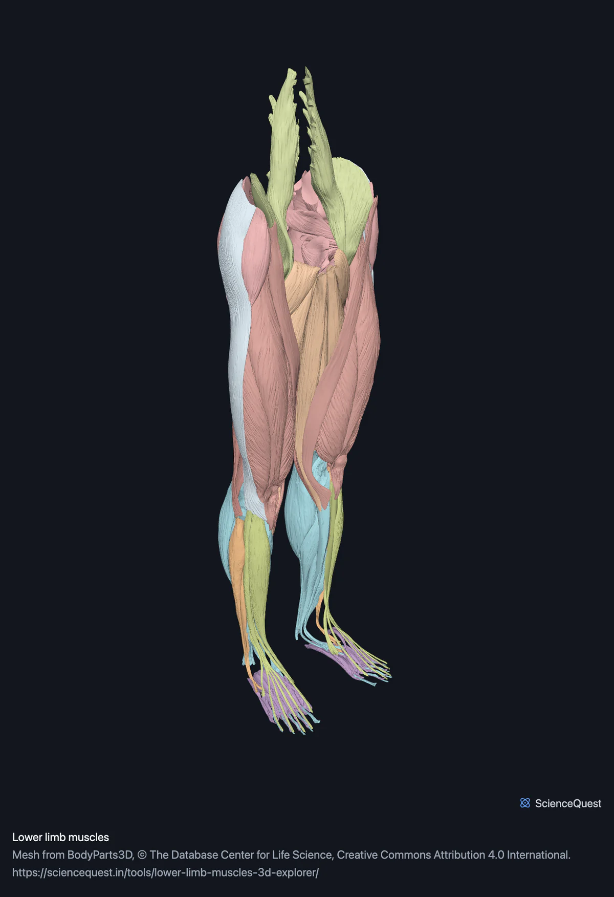
Turn 122 muscle meshes of both lower limbs in 3D, banded by compartment: gluteals, quadriceps, adductors, hamstrings, calf and the foot.
Visualiser
Lower limb muscles, 126 structures
Checking this device for a stored copy.
Start here
- Hide the Foot band first It is 40 of the 126 meshes, all of them small and buried, so hiding it takes about a third of the structures off the screen, though under a tenth of the triangles, and costs you nothing until you want the intrinsics.
- Hide Anterior thigh, then click Medial thigh The adductors fill the inner thigh, medial to the quadriceps, and from the front only part of the group shows, mainly the pectineus, adductor longus and gracilis: the brevis and most of the magnus lie deeper. This is the pair of clicks that exposes them as a group.
- Click Posterior thigh, then Back Frames the hamstrings from behind, where the split between the long and short heads of the biceps femoris actually reads.
Muscle, and the calcaneal tendons and iliotibial tracts. No bone, no nerves, no vessels and no joint capsules: those are separate models or absent from the dataset. Individual fascicles are not separable, and neither are the tendons of most muscles, which are part of the belly mesh rather than their own.
- 126 structures
- 4597k triangles
- 23700 KB, on request
- 0.00849 mm worst error per axis
- 20 gluteal and deep hip
- 4 iliopsoas
- 10 anterior thigh
- 12 medial thigh
- 8 posterior thigh
- 8 anterior leg
- 4 lateral leg
- 18 posterior leg
- 40 foot
- 2 fascia and tendon
Where this comes from
Segmented from imaging of one adult male body, not drawn by an artist, so the shapes are measurements. Two consequences worth knowing: it is one person’s anatomy rather than an average, and faint stepping on some surfaces is the resolution of the original scan rather than a rendering fault.
Meshes from BodyParts3D, © The Database Center for Life Science, under Creative Commons Attribution 4.0 International. Names and identifiers are the dataset’s own: Foundational Model of Anatomy identifiers and English labels, as its parts list gives them. Converted meshes are shared under the same licence.
Changes made: Meshes were converted from binary STL to an indexed mesh with 16-bit quantised vertex positions, and surface normals were recomputed from the geometry. No vertex was moved other than by that quantisation, whose worst-case error on any one coordinate is reported on the page.
Latin terms for 86 of the 126 structures come from English Wikipedia anatomy infoboxes, under Creative Commons Attribution-ShareAlike 4.0, joined by Wikidata property P1402, Foundational Model of Anatomy identifier. Only a term a source states for that structure is used, never one constructed from a family name, which is why the rest carry no Latin rather than a guess.
All 126 structures
Every structure the model contains, group by group and alphabetically within each. Each name opens the viewer with that structure selected, so a link can point at one structure rather than at the whole region.
Gluteal and deep hip 20
- left gemellus inferior muscle
- left gemellus superior muscle
- left gluteus maximus musculus gluteus maximus muscle
- left gluteus medius musculus gluteus medius muscle
- left gluteus minimus musculus gluteus minimus muscle
- left obturator externus musculus obturatorius externus muscle
- left obturator internus musculus obturatorius internus muscle
- left piriformis musculus piriformis muscle
- left quadratus femoris musculus quadratus femoris muscle
- left tensor fasciae latae musculus tensor fasciae latae muscle
- right gemellus inferior muscle
- right gemellus superior muscle
- right gluteus maximus musculus gluteus maximus muscle
- right gluteus medius musculus gluteus medius muscle
- right gluteus minimus musculus gluteus minimus muscle
- right obturator externus musculus obturatorius externus muscle
- right obturator internus musculus obturatorius internus muscle
- right piriformis musculus piriformis muscle
- right quadratus femoris musculus quadratus femoris muscle
- right tensor fasciae latae musculus tensor fasciae latae muscle
Iliopsoas 4
- left iliacus musculus iliacus muscle
- left psoas major musculus psoas major muscle
- right iliacus musculus iliacus muscle
- right psoas major musculus psoas major muscle
Anterior thigh 10
- left rectus femoris musculus rectus femoris muscle
- left sartorius musculus sartorius muscle
- left vastus intermedius musculus vastus intermedius muscle
- left vastus lateralis musculus vastus lateralis, musculus vastus externus muscle
- left vastus medialis musculus vastus medialis, musculus vastus internus muscle
- right rectus femoris musculus rectus femoris muscle
- right sartorius musculus sartorius muscle
- right vastus intermedius musculus vastus intermedius muscle
- right vastus lateralis musculus vastus lateralis, musculus vastus externus muscle
- right vastus medialis musculus vastus medialis, musculus vastus internus muscle
Medial thigh 12
- left adductor brevis musculus adductor brevis muscle
- left adductor longus musculus adductor longus muscle
- left adductor magnus musculus adductor magnus muscle
- left adductor minimus musculus adductor minimus muscle
- left gracilis musculus gracilis muscle
- left pectineus musculus pectineus muscle
- right adductor brevis musculus adductor brevis muscle
- right adductor longus musculus adductor longus muscle
- right adductor magnus musculus adductor magnus muscle
- right adductor minimus musculus adductor minimus muscle
- right gracilis musculus gracilis muscle
- right pectineus musculus pectineus muscle
Posterior thigh 8
- left semimembranosus musculus semimembranosus muscle
- left semitendinosus musculus semitendinosus muscle
- long head of left biceps femoris muscle
- long head of right biceps femoris muscle
- right semimembranosus musculus semimembranosus muscle
- right semitendinosus musculus semitendinosus muscle
- short head of left biceps femoris muscle
- short head of right biceps femoris muscle
Anterior leg 8
- left extensor digitorum longus musculus extensor digitorum longus muscle
- left extensor hallucis longus musculus extensor hallucis longus muscle
- left fibularis tertius musculus fibularis tertius muscle
- left tibialis anterior musculus tibialis anterior muscle
- right extensor digitorum longus musculus extensor digitorum longus muscle
- right extensor hallucis longus musculus extensor hallucis longus muscle
- right fibularis tertius musculus fibularis tertius muscle
- right tibialis anterior musculus tibialis anterior muscle
Lateral leg 4
- left fibularis brevis musculus fibularis brevis muscle
- left fibularis longus musculus fibularis longus muscle
- right fibularis brevis musculus fibularis brevis muscle
- right fibularis longus musculus fibularis longus muscle
Posterior leg 18
- lateral head of left gastrocnemius muscle
- lateral head of right gastrocnemius muscle
- left calcaneal tendon tendo calcaneus, tendo Achillis other tissue
- left flexor digitorum longus musculus flexor digitorum longus muscle
- left flexor hallucis longus musculus flexor hallucis longus muscle
- left plantaris musculus plantaris muscle
- left popliteus muscle
- left soleus musculus soleus muscle
- left tibialis posterior musculus tibialis posterior muscle
- medial head of left gastrocnemius muscle
- medial head of right gastrocnemius muscle
- right calcaneal tendon tendo calcaneus, tendo Achillis other tissue
- right flexor digitorum longus musculus flexor digitorum longus muscle
- right flexor hallucis longus musculus flexor hallucis longus muscle
- right plantaris musculus plantaris muscle
- right popliteus muscle
- right soleus musculus soleus muscle
- right tibialis posterior musculus tibialis posterior muscle
Foot 40
- abductor digiti minimi of left foot musculus abductor digiti minimi pedis muscle
- abductor digiti minimi of right foot musculus abductor digiti minimi pedis muscle
- first lumbrical of left foot muscle
- first lumbrical of right foot muscle
- first plantar interosseous of left foot muscle
- first plantar interosseous of right foot muscle
- flexor digiti minimi brevis of left foot musculus flexor digiti minimi brevis pedis muscle
- flexor digiti minimi brevis of right foot musculus flexor digiti minimi brevis pedis muscle
- fourth lumbrical of left foot muscle
- fourth lumbrical of right foot muscle
- lateral head of left flexor hallucis brevis muscle
- lateral head of right flexor hallucis brevis muscle
- left abductor hallucis musculus abductor hallucis muscle
- left extensor digitorum brevis musculus extensor digitorum brevis muscle
- left extensor hallucis brevis musculus extensor hallucis brevis muscle
- left flexor accessorius musculus quadratus plantae muscle
- left flexor digitorum brevis musculus flexor digitorum brevis muscle
- medial head of left flexor hallucis brevis muscle
- medial head of right flexor hallucis brevis muscle
- oblique head of left adductor hallucis muscle
- oblique head of right adductor hallucis muscle
- opponens digiti minimi of left foot muscle
- opponens digiti minimi of right foot muscle
- right abductor hallucis musculus abductor hallucis muscle
- right extensor digitorum brevis musculus extensor digitorum brevis muscle
- right extensor hallucis brevis musculus extensor hallucis brevis muscle
- right flexor accessorius musculus quadratus plantae muscle
- right flexor digitorum brevis musculus flexor digitorum brevis muscle
- second lumbrical of left foot muscle
- second lumbrical of right foot muscle
- second plantar interosseous of left foot muscle
- second plantar interosseous of right foot muscle
- set of dorsal interossei of left foot muscle
- set of dorsal interossei of right foot muscle
- third lumbrical of left foot muscle
- third lumbrical of right foot muscle
- third plantar interosseous of left foot muscle
- third plantar interosseous of right foot muscle
- transverse head of left adductor hallucis muscle
- transverse head of right adductor hallucis muscle
Fascia and tendon 2
- left iliotibial tract tractus iliotibialis other tissue
- right iliotibial tract tractus iliotibialis other tissue
Citing this tool
Last updated . Add the date you accessed it as well, which a citation of a page that can change asks for. If a specific view matters, cite the page address as it stands in your browser instead: it records what is selected and shown.
The heaviest model here, and why it ships anyway
This is 4.6 million triangles against 2.36 million for the entire skeleton. Muscle is the expensive kind of geometry: bulky smooth volumes with no flat faces to collapse, and there are 122 of them, 126 structures with the tendons and tracts. Compressed it is about 23 MB.
It ships at full resolution rather than being trimmed to a number somebody else chose. Nothing is fetched until you press the load button, the figure is printed on that button, and once loaded it is kept on your device with a button to remove it again. A download you asked for is your decision to make.
The limit that is not a matter of preference is graphics memory. A scene this size needs roughly 90 MB before anything is drawn, which a laptop has and an older phone may not. If the device refuses, the viewer falls back to the structure list rather than to an error, so all 126 names are still there to search. Hiding a band does not reduce that figure, because a hidden mesh keeps its buffers in graphics memory.
Bands are compartments, because that is where the information is
The medial thigh muscles share a nerve and an action. So do the hamstrings, and so does each group in the leg. A clinician thinks in compartments rather than in individual bellies, and compartment syndrome is named for the fact. Grouping this way also makes the model usable: hiding the anterior thigh to see the adductors behind it is one click instead of five.
Four of the 126 are not muscle
The calcaneal tendon, which is the Achilles, is tendon, and the iliotibial tract is fascia, one of each per side. Both are tagged as other tissue and left out of the muscle count, which is why 122 of the 126 structures are muscle meshes.
They are in the model because they are what the muscles pull through. A calf without the Achilles ends in mid air, and a lateral thigh without the tract loses the sheet that the tensor fasciae latae and part of the gluteus maximus actually insert into.
Mapped by name, checked against the dataset
126 identifiers is too many to transcribe safely, and transcription is exactly where a part goes missing without anything failing. So the bands name muscles rather than ids, the build strips the side word to match them, and it then asserts that every leaf under the dataset’s lower limb muscle root found a band and that no band names a muscle which is not there. A typo fails the build instead of quietly shipping 125.
Common questions
Why is this the heaviest model here?
Because it is 4.6 million triangles, against 2.36 million for the whole skeleton. Muscles are bulky smooth volumes and there are 122 muscle meshes, 126 structures with the tendons and tracts, where a skeleton is mostly thin hard shells. It comes to about 23 MB compressed, and it ships at full resolution rather than being trimmed: the figure is printed on the load button, nothing is fetched until you press it, and it is kept on your device afterwards with a button to remove it. The one thing worth knowing before you press it is the graphics memory, not the download.
Will it run on my device?
On a laptop or desktop, yes. On a phone it may not: a scene this size needs roughly 90 MB of graphics memory before anything is drawn, and an older phone will refuse that. If it does refuse, the viewer falls back to the structure list rather than showing an error, so you still get all 126 names and can still search them. Hiding a band does not reduce that 90 MB, because a hidden mesh keeps its buffers in graphics memory.
Why bands by compartment rather than by joint?
Because the compartment is the unit that carries the information. The muscles of the medial thigh share a nerve and an action, the hamstrings share another, and a surgeon or a physiotherapist thinks in those groups rather than in individual bellies. It also makes the model usable: hiding the anterior thigh to see the adductors behind it is one click, where hiding five muscles individually is five.
Is anything in here not a muscle?
Two things, and they are tagged as other tissue so they are not counted among the muscles. The calcaneal tendon, the Achilles, is tendon rather than muscle, and the iliotibial tract is fascia. Both are in the model because they are what the muscles pull through, and leaving them out would make the calf and the lateral thigh read wrongly. That is why the count says 122 muscle meshes rather than 126 structures.