Chest Wall Muscles Explorer
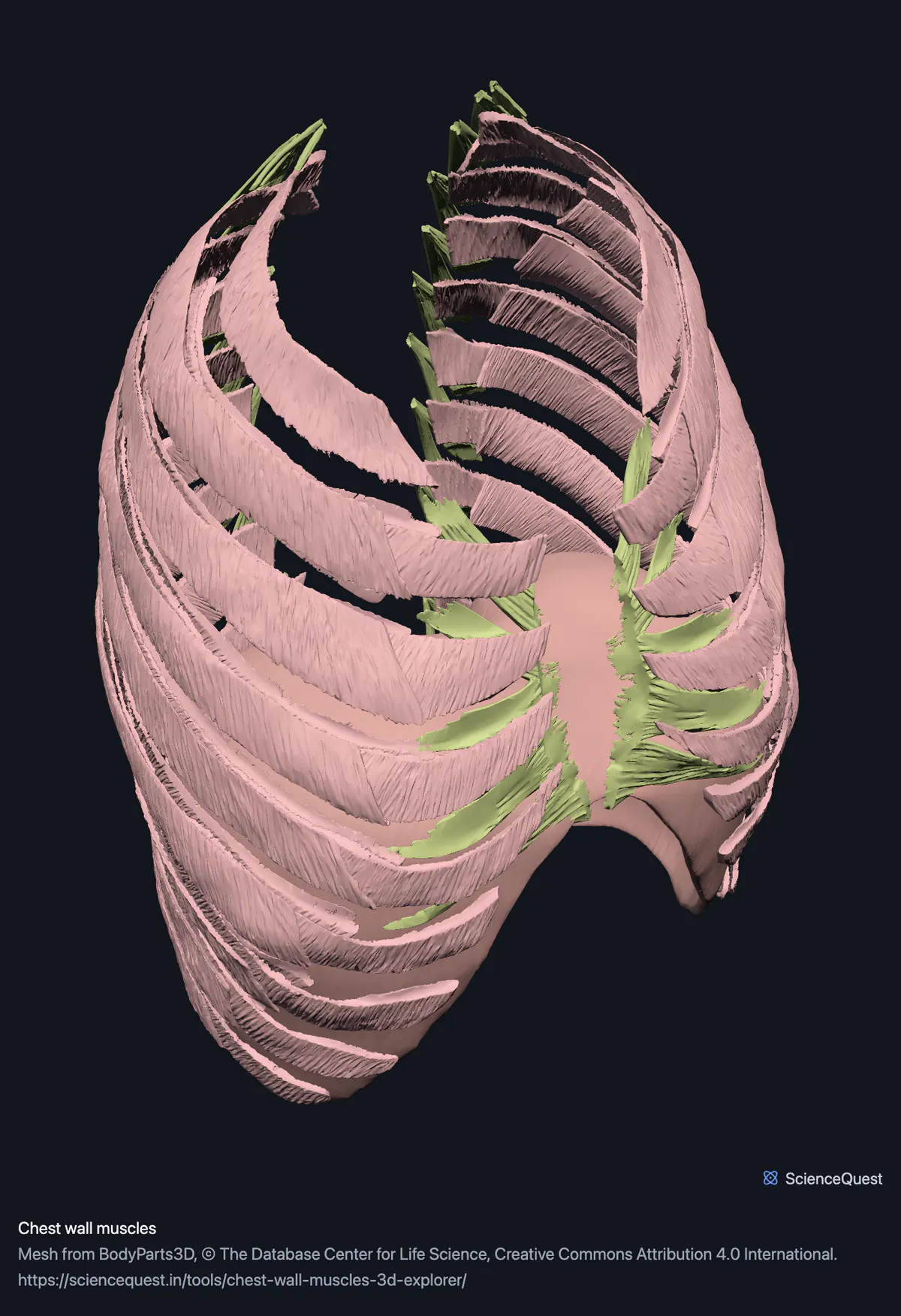
Turn the chest wall in 3D: the diaphragm as a single dome, all three intercostal layers, the levatores costarum and transversus thoracis.
Visualiser
Chest wall muscles, 10 structures
Checking this device for a stored copy.
Start here
- Click Diaphragm twice, then Front Opening a band frames it, and this one starts open, so the second click is the one that takes you there. One mesh, and the thing most diagrams get wrong. It reaches far higher into the chest than a flat drawing suggests, and the two domes are not level with each other.
- Hide the Intercostals Three meshes hide the inner surface of the whole rib cage, including transversus thoracis, which is the one chest wall muscle almost nobody sees.
- Turn on Cut plane and drag it Sections the wall so the three intercostal layers read as three, which from outside they never do.
Muscle only. No ribs, no pleura and no sternum: the rib cage is a separate model. The intercostals cannot be separated space by space, because each layer is published as one mesh for the whole thorax.
- 10 structures
- 2639k triangles
- 13000 KB, on request
- 0.00308 mm worst error per axis
- 1 diaphragm
- 3 intercostals
- 6 levatores and transversus
Where this comes from
Segmented from imaging of one adult male body, not drawn by an artist, so the shapes are measurements. Two consequences worth knowing: it is one person’s anatomy rather than an average, and faint stepping on some surfaces is the resolution of the original scan rather than a rendering fault.
Meshes from BodyParts3D, © The Database Center for Life Science, under Creative Commons Attribution 4.0 International. Names and identifiers are the dataset’s own: Foundational Model of Anatomy identifiers and English labels, as its parts list gives them. Converted meshes are shared under the same licence.
Changes made: Meshes were converted from binary STL to an indexed mesh with 16-bit quantised vertex positions, and surface normals were recomputed from the geometry. No vertex was moved other than by that quantisation, whose worst-case error on any one coordinate is reported on the page.
Latin terms for 4 of the 10 structures come from English Wikipedia anatomy infoboxes, under Creative Commons Attribution-ShareAlike 4.0, joined by Wikidata property P1402, Foundational Model of Anatomy identifier. Only a term a source states for that structure is used, never one constructed from a family name, which is why the rest carry no Latin rather than a guess.
All 10 structures
Every structure the model contains, group by group and alphabetically within each. Each name opens the viewer with that structure selected, so a link can point at one structure rather than at the whole region.
Diaphragm 1
- diaphragm diaphragma muscle
Intercostals 3
- external intercostal muscle muscle
- innermost intercostal muscle muscle
- internal intercostal muscle musculi intercostales interni muscle
Levatores and transversus 6
- left transversus thoracis musculus transversus thoracis muscle
- right transversus thoracis musculus transversus thoracis muscle
- set of left levatores costarum breves muscle
- set of left levatores costarum longi muscle
- set of right levatores costarum breves muscle
- set of right levatores costarum longi muscle
Citing this tool
Last updated . Add the date you accessed it as well, which a citation of a page that can change asks for. If a specific view matters, cite the page address as it stands in your browser instead: it records what is selected and shown.
Ten meshes, and more triangles than the skeleton
This region has 2.64 million triangles spread across ten objects, where the full skeleton has 2.36 million spread across 248. The reason is how the dataset divides it: each intercostal layer is a single mesh covering every space on both sides at once, and the diaphragm is one broad dome.
The consequence is worth knowing before you use it. You can show or hide the external layer as a whole, and you cannot isolate the fourth left intercostal space, because it is not a separate object in the data.
What it shows about breathing, and what it cannot
Nothing here moves. These are static surfaces from one instant of one body, so the diaphragm sits wherever that person’s was at the moment of imaging.
What the geometry does show is why the mechanism works. The external and internal intercostals run in opposite directions, which is the entire basis of one raising the ribs and the other lowering them, and fibre direction is far clearer on a curved surface you can turn than on a printed diagram of two flat hatched panels.
The pectorals are in the upper limb model
Pectoralis major, pectoralis minor, serratus anterior and subclavius sit under this root as well, and they ship with the limb they move. That is what keeps this region the wall itself, and it is also what lets the intercostals be seen from the front: with the pectoral group shown, most of the upper front and side of the chest wall is covered.
Common questions
Why only ten structures for a whole chest wall?
Because of how the dataset divides it, and the ratio is the strangest here: ten meshes carrying 2.64 million triangles, which is more than the entire skeleton. Each intercostal layer is published as a single mesh covering every space on both sides at once, and the diaphragm is one broad dome. So you get the external, internal and innermost layers as three objects rather than as sixty six separate sheets, one per layer in each of the twenty two spaces, and no amount of clicking will separate one intercostal space from the next.
Can I see how breathing works?
You can see the machinery, not the movement. Nothing here animates: these are static surfaces from one instant of one body, so the diaphragm is captured at whatever position that person’s was. What the model does show is why the geometry works, which a diagram tends to flatten: the dome shape, how far up into the chest the diaphragm reaches, and the opposite fibre directions of the external and internal intercostals, which is the whole basis of one raising the ribs and the other lowering them.
Where are the pectorals?
In the upper limb model. The dataset files pectoralis major, pectoralis minor, serratus anterior and subclavius under the thorax as well as the upper limb, and they ship with the limb because what they do is move the arm. That keeps this region the wall itself rather than the wall plus the muscles lying on top of it, which is also what keeps the intercostals visible from the front.
Are the diaphragm’s openings there?
The shape of them, yes, since they are holes in a surface rather than separate objects: you can find where the aorta, the oesophagus and the inferior vena cava pass through. What is not here is anything passing through them. Open the great vessels model alongside this one and they line up, because every region here shares one coordinate frame.