DNA Double Helix Explorer
Turn a double helix and see the major and minor grooves, which exist only because the two backbones are not directly opposite each other.
Visualiser
Drag the helix to turn it, or use the arrow keys. Space plays and pauses.
- Base pairs per turn 34.3 degrees of twist per pair, so 360 divided by that.
- 10.5
- Pitch Length of one full turn. Rise per pair times pairs per turn.
- 3.57 nm
- Major groove The wider gap between the backbones. The narrow one is 120 degrees.
- 240 deg
- Handedness Right handed, like almost all DNA in a cell.
- right
- GC content 52 hydrogen bonds across 21 pairs.
- 47.6 %
- Melting temperature Wallace rule, outside the 14 to 20 base range it was derived for, so treat it as indicative.
- 62 deg C
Both strands, each read 5′ to 3′ ATGGCATTACGCTAGCATGCA TGCATGCTAGCGTAATGCCAT
The lower strand is the complement written backwards, because the two run in opposite directions. Reading it forwards is the commonest slip: it produces a sequence that looks right and pairs with nothing.
- B-DNA The form in living cells. Ten and a half base pairs per turn, and a major groove wide enough for a protein to read the bases.
- A-DNA Dehydrated DNA, and the shape RNA duplexes take. Shorter rise and a fatter helix, so it is more compressed along its length.
- Z-DNA Left handed, found in alternating purine and pyrimidine tracts. Its zigzag backbone leaves almost no major groove.
Citing this tool
Last updated . Add the date you accessed it as well, which a citation of a page that can change asks for. If a specific result matters, cite the permalink from the tool’s share row instead of this page: it reproduces the exact parameters.
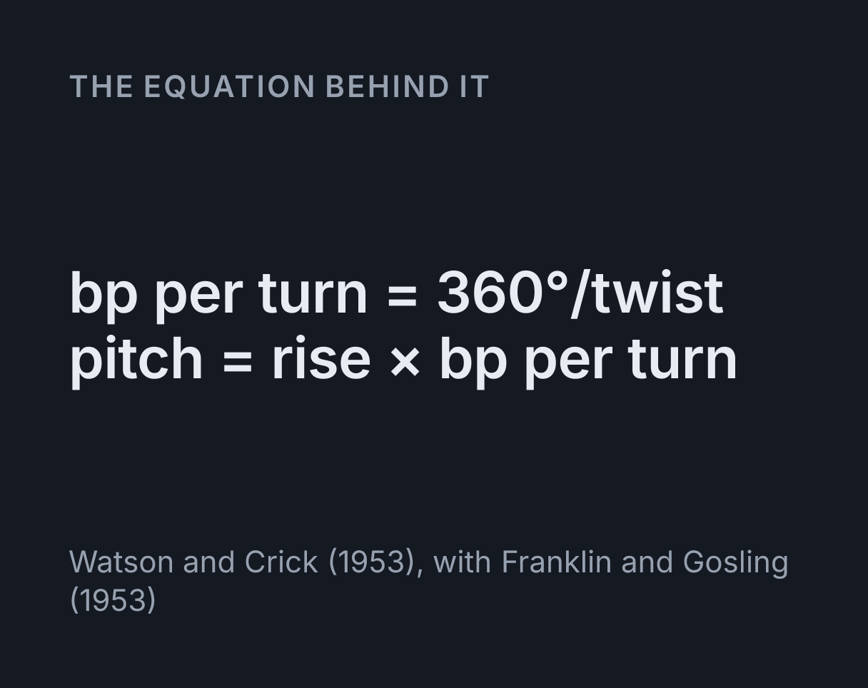
The equation
Watson and Crick (1953), with Franklin and Gosling (1953)
The thing almost every diagram gets wrong
Look at a textbook drawing of DNA and the two backbones will be directly opposite each other, half a turn apart. If that were true, the two gaps between them would be identical, and there would be no major groove and no minor groove.
There are two grooves, of quite different widths, and the reason is that the base pair does not pass through the axis of the helix. It joins the two backbones at points about 120 degrees apart, not 180. One gap therefore covers roughly 240 degrees of the circle and the other roughly 120.
That single asymmetry is the entire explanation. Turn the model slowly with the base pairs hidden and watch the wide gap and the narrow one alternate as they come round.
Why the wide groove is the one that matters
Because only it carries enough information to tell the base pairs apart. The edges of the bases facing into the major groove differ enough between the four possible pairs that a protein touching them can identify which is which. The edges facing the minor groove are much more alike.
So most sequence-specific DNA binding proteins, including the great majority of transcription factors, read the major groove. Proteins and drugs that bind the minor groove tend to recognise shape and flexibility instead, which is why several minor-groove binders prefer AT-rich stretches without reading a specific word.
Three numbers, and only two of them are independent
B-DNA rises 0.34 nm per base pair and twists about 34.3 degrees per pair. Everything else follows.
| Quantity | Value | Where from |
|---|---|---|
| Base pairs per turn | 10.5 | 360 ÷ 34.3 |
| Pitch | 3.6 nm | 0.34 × 10.5 |
| Diameter | 2.0 nm | Measured |
The pitch is not a third fact to memorise. The model derives it, so changing the twist moves it correctly rather than needing a second table that could disagree.
Antiparallel, and the error it causes
Each strand has a chemical direction, read from its 5′ end to its 3′ end, and the two strands of a duplex point opposite ways. So to write a complement in the same 5′ to 3′ convention, you have to reverse it.
The complement of ATGC is GCAT, not
TACG. Writing it forwards is the commonest slip in the topic
and it produces a sequence that looks entirely plausible and pairs with
nothing. Both strands are printed under the model so you can check.
To turn a whole sequence round, ambiguity codes and all, paste it into the reverse complement calculator, which labels the direction every result reads in.
One consequence worth noticing: a sequence like GAATTC reads
identically on both strands. That is what a palindromic restriction site
is, and it is why enzymes that cut as a pair of identical subunits
recognise sequences of that shape.
Why GC-rich DNA is harder to melt
A G to C pair has three hydrogen bonds where A to T has two, so it takes more energy to pull apart. The rungs in the model are coloured by bond count, so a GC-rich stretch looks different from an AT-rich one.
The part usually left out is that stacking matters more than the hydrogen bonds. Neighbouring bases interact face to face, GC steps stack more strongly, and that contributes more to holding the duplex together than the bonds across it do.
The melting temperature shown uses the Wallace rule, four degrees per G or C and two per A or T. It is crude, it is what gets used at the bench for short primers, and it is only valid for roughly 14 to 20 bases. The readout says so when your sequence falls outside that. For a primer’s Tm with salt, magnesium and base order taken into account, use the primer Tm calculator.
The other two forms
A-DNA appears when DNA is dehydrated, and it is the shape RNA duplexes adopt. Still right handed, but with a shorter rise and a fatter helix, so it is compressed along its length.
Z-DNA is the interesting one: left handed, found in alternating purine and pyrimidine tracts. Its zigzag backbone leaves the surface almost uniform, so it has no major groove worth the name, and the model says that rather than drawing a groove that is not there. Its handedness comes from a negative twist, so the helix winds the other way as arithmetic rather than as a special case.
Common mistakes
- Drawing the strands directly opposite. That removes both grooves, which is most of what the structure does.
- Writing the complement without reversing it. The strands
are antiparallel.
ATGCpairs withGCAT. - Saying 10 base pairs per turn. It is about 10.5, which is why 21 base pairs is a convenient two full turns.
- Thinking hydrogen bonds alone hold the duplex together. Base stacking contributes more. The bonds give the specificity; the stacking gives much of the stability.
- Assuming all DNA is right handed. Z-DNA is left handed and occurs in real sequences.
- Trusting the Wallace rule outside its range. Below about 14 bases or above 20, salt concentration and stacking dominate and the base counts stop being a good guide.
Common questions
Why does DNA have a major groove and a minor groove?
Because the two backbones are not directly opposite each other. Almost every diagram draws them half a turn apart, and if that were true the two gaps between them would be identical and there would be no major or minor groove at all. In reality the base pair does not pass through the helix axis, so the two points where it joins the backbones sit about 120 degrees apart rather than 180. That leaves one gap subtending roughly 240 degrees of the circle and the other roughly 120, which is the wide groove and the narrow one. Turning the model slowly is the clearest way to see it, because the two gaps alternate as they come round.
Why does it matter which groove a protein binds in?
Because only one of them carries enough information to identify the base pairs. The edges of the bases that face into the major groove differ enough between the four possible pairs that a protein touching them can tell which is which, while the edges facing the minor groove are much more alike. That is why the great majority of sequence-specific DNA binding proteins, including most transcription factors, read the major groove. Proteins that bind the minor groove tend to recognise shape and flexibility rather than sequence, which is also why several minor-groove-binding drugs prefer AT-rich stretches without reading a specific word.
What is the difference between B-DNA, A-DNA and Z-DNA?
B-DNA is the form found in cells under normal conditions and is what people mean by DNA: right handed, about 10.5 base pairs per turn, 0.34 nm rise per pair and 2 nm across. A-DNA appears when DNA is dehydrated and is the shape RNA duplexes adopt: still right handed but shorter in rise and fatter, so it is compressed along its length. Z-DNA is the odd one, a left handed helix found in alternating purine and pyrimidine tracts, whose zigzag backbone leaves the surface almost uniform so it has no major groove worth the name. Switching between them here changes the geometry, and the handedness of Z follows from a negative twist rather than from a label.
Why must the complementary strand be written backwards?
Because the two strands run in opposite directions, which is what antiparallel means. Each strand has a chemical direction, conventionally read from its 5′ end to its 3′ end, and the two strands of a duplex point opposite ways. So if you write one strand 5′ to 3′ and want to write its partner the same way, you have to reverse it. The complement of ATGC read 5′ to 3′ is GCAT, not TACG. Writing the complement without reversing it is the single commonest error in this topic, and the result looks entirely plausible while pairing with nothing.
Why do GC-rich sequences melt at a higher temperature?
Two reasons, and the second is often left out. A guanine and cytosine pair is held by three hydrogen bonds where adenine and thymine manage two, so more energy is needed to separate it. Just as importantly, the stacking interactions between neighbouring bases are stronger for GC steps, and stacking contributes more to duplex stability than the hydrogen bonds do. The Wallace rule used here counts four degrees per G or C and two per A or T, which captures the effect crudely and is what people use at the bench for short primers. It is only valid for roughly 14 to 20 bases, and the readout says when your sequence is outside that range.
How long is a turn of DNA?
About 3.6 nanometres for B-DNA, which follows from the two numbers everyone learns rather than being a third fact to remember: 0.34 nm of rise per base pair times roughly 10.5 base pairs per turn. That is why the model derives pitch from rise and twist rather than storing it, so changing the twist moves the pitch correctly. For a sense of scale, the DNA in one human cell is around two metres long and has to fit inside a nucleus about six micrometres across, which is the reason chromatin packing exists.