Muscles Worked by Exercise 3D Explorer
Pick a lift, from the squat and deadlift to the pull-up, and see the muscles it works on a real 3D body, coloured as prime mover, synergist or stabiliser.
Visualiser
Squat
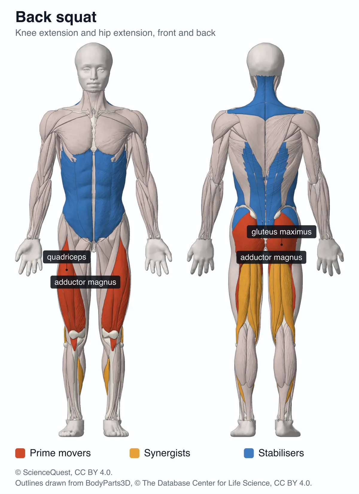
Back squat
Knee extension and hip extension
Rising: they shorten to extend the knees and hips
Point at or tap a muscle to see its role in this lift.
Under other muscles here: multifidus, internal oblique, transversus abdominis, quadratus lumborum, gluteus minimus and rhomboids.
Assumed: Barbell high on the upper back, shoulder-width stance, lowered until the thighs are parallel to the floor.
Sources: Escamilla (2001), Med Sci Sports Exerc 33(1):127; Schoenfeld (2010), J Strength Cond Res 24(12):3497; Németh and Ohlsén (1985), J Biomech 18(2):129; NSCA, Essentials of Strength Training and Conditioning, 4th ed. (2016).
The same muscles in 3D
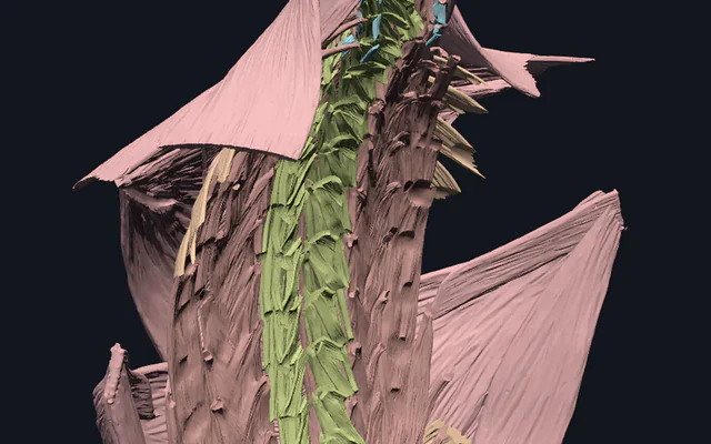
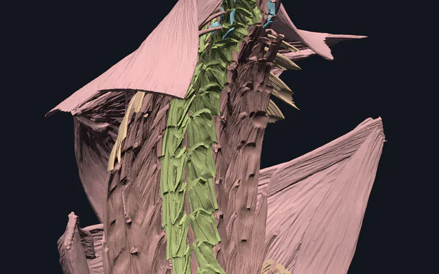
168 meshes from the BodyParts3D muscle models, coloured as the map is, with every muscle the lift does not use turned to glass. The camera frames the lift’s muscles each time you change it.
Muscles worked by exercise, 168 structures
The model is about 18.0 MB and is not downloaded until you ask for it, so this page stays fast for everyone else. It is kept on your device afterwards, so this is a one-off.
18.0 MB · segmented anatomical imaging
Start here
- Turn off Stabilisers and Synergists The map keeps only the prime movers, the muscles that produce the lift’s main joint action, and then adds the helpers back one role at a time.
- Tap a muscle, then Exercises for this muscle Every lift here that works it, ranked by role: the gluteus maximus is a prime mover in 9 of the 25.
- Tick Deep muscles as outlines Muscles that lie under others, such as the vastus intermedius and gluteus minimus, are drawn as dashed outlines through the ones above them.
Muscle, from five of the six BodyParts3D muscle models, with the calcaneal tendons. No bone, nerves, vessels or skin in the 3D model, and none of the hand, foot or face muscles except the one hand muscle the myotome table needs. The flat map draws bone and skin for context only. Roles are for the stated technique of each lift and are not measurements of how hard a muscle works.
- 168 structures
- 4893k triangles
- 18044 kB, on request
- 0.0200 mm worst error per axis
- 4 neck
- 10 chest
- 16 shoulder
- 14 arm
- 34 forearm and hand
- 26 back
- 8 abdominal wall
- 12 hip
- 28 thigh
- 16 leg
Simplified from 10,388,058 triangles to 4,893,458, and no original vertex lies more than 0.667 mm from the simplified surface, measured mesh by mesh. One mesh ships whole, because no tolerance kept it within that: the right external oblique.
Where this comes from
Segmented from MRI of one adult male volunteer, then refined by medical illustrators, who filled in missing detail and sharpened blurred outlines from textbooks and atlases. Two consequences worth knowing: it is one person’s anatomy rather than an average, and faint stepping on some surfaces is the resolution of the original scan rather than a rendering fault.
Meshes from BodyParts3D, © The Database Center for Life Science, under Creative Commons Attribution 4.0 International. Names and identifiers are the dataset’s own: Foundational Model of Anatomy identifiers and English labels, as its parts list gives them. Converted meshes are shared under the same licence.
Changes made: Meshes were converted from binary STL to an indexed mesh with 16-bit quantised vertex positions, and surface normals were recomputed from the geometry. No vertex was moved other than by that quantisation, whose worst-case error on any one coordinate is reported on the page. This model was then made from a selection of those converted meshes: each was simplified by edge collapse that keeps only original vertices, at the largest tolerance whose measured distance from every original vertex to the simplified surface stays within 0.7 mm, or left whole where none did, and its positions were quantised again to 16 bits over this model’s own bounds. Each model’s file is then compressed without loss, by meshoptimizer’s vertex and index codecs and gzip, and restored exactly before it is drawn.
Latin terms for 108 of the 168 structures come from English Wikipedia anatomy infoboxes, under Creative Commons Attribution-ShareAlike 4.0, joined by Wikidata property P1402, Foundational Model of Anatomy identifier. Only a term a source states for that structure is used, never one constructed from a family name, which is why the rest carry no Latin rather than a guess.
Citing this tool
Last updated . Add the date you accessed it as well, which a citation of a page that can change asks for. If a specific view matters, cite the page address as it stands in your browser instead: it records what is selected and shown.
What muscles are activated during compound strength exercises?
Compound exercises move more than one joint, so several muscles share the work. The prime movers of the back squat are the quadriceps, gluteus maximus and adductor magnus; of the bench press, pectoralis major, the anterior deltoid and triceps brachii; and of the deadlift, gluteus maximus, the hamstrings, adductor magnus, the quadriceps and the erector spinae, which hold the spine still.
Prime mover, synergist, stabiliser: how a role is decided here
Every lift here is described joint by joint. First come the main joint actions the lift is made of, such as knee extension and hip extension in a squat. Then each muscle gets the one role that fits what it does in that technique:
- Prime mover. It produces the lift’s main joint action against the load.
- Synergist. It assists that action or adds a secondary one.
- Stabiliser. It contracts mostly isometrically to hold a joint or segment still.
A prime mover always sits under one of the lift’s joint actions, and no muscle has two roles in one lift. Where a prime mover works without changing length, the card says so: in the deadlift the erector spinae are prime movers that hold the spine still rather than move it. The roles follow kinesiology texts (Neumann; Kendall and colleagues; the NSCA’s Essentials of Strength Training and Conditioning) and the electromyography studies named on each lift, which settle how two variations differ: that a chin-up uses more biceps and a pull-up more lower trapezius (Youdas and colleagues, 2010), or that a sumo deadlift uses more of the vasti (Escamilla and colleagues, 2002).
Worked example: the back squat joint by joint
The back squat opens the page, done this way: barbell high on the upper back, shoulder-width stance, lowered until the thighs are parallel to the floor.
- Knee extension. The quadriceps, all four heads, straighten the knee out of the bottom against the load, and that joint action is what makes them prime movers. They lengthen under load on the way down and shorten on the way up.
- Hip extension. Gluteus maximus and adductor magnus drive the hips forward and up. The hamstrings help, but they also cross the knee, which straightens as the hip does; they lengthen at the knee as they shorten at the hip, so their length changes much less than that of gluteus maximus: synergists.
- The ankle. Soleus controls how far the shin tips forward, a synergist.
- Everything held still. Erector spinae, multifidus, rectus abdominis, external oblique, internal oblique, transversus abdominis, quadratus lumborum, gluteus medius, gluteus minimus, upper trapezius, middle trapezius and rhomboids: the trunk, the pelvis and the shoulder blades the bar rests on, which do their work without moving.
That is 3 prime movers, 2 synergists and 12 stabilisers, the counts beside the map. Going deeper or widening the stance moves more of the work to the hip extensors (Escamilla, 2001; Schoenfeld, 2010), which a role cannot show and the card notes where it matters.
Why muscle charts disagree
Two charts of the same lift often light different muscles, and both can be right, because each assumes a technique. Depth, stance width, grip, bar position, tempo and load all shift the work between muscles, and so do the lifter’s proportions. That is why every lift here states the technique its roles are for, under the map.
Charts are also often read as rankings of growth, which they are not. Surface electromyography measures how strongly a muscle is activated, and the signal depends on where the electrodes sit, how it was normalised and the person; a larger amplitude does not necessarily mean more force or more growth (Vigotsky et al. (2018), Front Physiol 8:985). So the studies cited here are used for which muscles a lift involves and how two variations differ, never to put a percentage on a muscle. How much a muscle grows depends mostly on its weekly volume and how close sets go to failure, and is much the same across a wide range of loads (Schoenfeld and colleagues, 2017), which no chart of a single lift can say.
Deep muscles the flat map cannot show
The maps are traced from the meshes as an orthographic camera sees them, so a muscle under another is hidden, as it is on a body. In both views the map cannot show any of the 10 muscles or muscle groups that lie wholly under others: levator scapulae, rhomboids, multifidus, quadratus lumborum, pectoralis minor, supraspinatus, subscapularis, supinator, internal oblique and gluteus minimus. Parts of others are hidden while the rest shows, such as the vastus intermedius under the other three heads of the quadriceps, and abductor digiti minimi of the hand is under the skin the map draws over the hands. Tick Deep muscles as outlines and every muscle the lift uses that is mostly hidden is drawn as a dashed outline of its whole shape, in its role’s colour, and the 3D model below shows each in place.
Three thin sheets are drawn as if transparent, because the meshes join each to its muscle. The external oblique carries its aponeurosis, which forms the front of the rectus sheath, so rectus abdominis would otherwise be hidden behind it, and is seen through to 15 mm. Latissimus dorsi carries the thoracolumbar fascia over the erector spinae, seen through to 20 mm, and platysma, a skin muscle, lies over sternocleidomastoid, seen through to 10 mm. Deeper than that, a muscle stays hidden.
Every lift at a glance
All 25 lifts, in the 9 movement patterns the picker uses, with the muscles in each role, from the same records the map is coloured from.
| Lift | Prime movers | Synergists | Stabilisers |
|---|---|---|---|
| Back squat | quadriceps; gluteus maximus; adductor magnus | hamstrings; soleus | erector spinae; multifidus; rectus abdominis; external oblique; internal oblique; transversus abdominis; quadratus lumborum; gluteus medius; gluteus minimus; upper trapezius; middle trapezius; rhomboids |
| Front squat | quadriceps; gluteus maximus; adductor magnus | hamstrings; soleus | erector spinae; multifidus; rectus abdominis; external oblique; internal oblique; transversus abdominis; gluteus medius; gluteus minimus; anterior deltoid; upper trapezius |
| Leg press | quadriceps; gluteus maximus; adductor magnus | hamstrings | soleus; gastrocnemius; gluteus medius; rectus abdominis; external oblique; internal oblique; transversus abdominis; wrist and finger flexors |
| Conventional deadlift | gluteus maximus; hamstrings; adductor magnus; quadriceps; erector spinae | gastrocnemius; soleus | multifidus; rectus abdominis; external oblique; internal oblique; transversus abdominis; quadratus lumborum; latissimus dorsi; upper trapezius; middle trapezius; lower trapezius; rhomboids; wrist and finger flexors; gluteus medius; tibialis anterior |
| Sumo deadlift | gluteus maximus; adductor magnus; hamstrings; quadriceps; erector spinae | adductor longus and brevis; gracilis | tibialis anterior; gastrocnemius; multifidus; rectus abdominis; external oblique; internal oblique; transversus abdominis; latissimus dorsi; upper trapezius; rhomboids; wrist and finger flexors; gluteus medius |
| Romanian deadlift | hamstrings; gluteus maximus; adductor magnus; erector spinae | none | quadriceps; gastrocnemius; multifidus; rectus abdominis; external oblique; internal oblique; transversus abdominis; latissimus dorsi; upper trapezius; rhomboids; wrist and finger flexors |
| Barbell hip thrust | gluteus maximus | hamstrings; adductor magnus; quadriceps | erector spinae; rectus abdominis; external oblique; internal oblique; transversus abdominis; gluteus medius; gluteus minimus |
| Bulgarian split squat | quadriceps; gluteus maximus; adductor magnus | hamstrings; soleus | gluteus medius; gluteus minimus; tensor fasciae latae; adductor longus and brevis; quadratus lumborum; erector spinae; rectus abdominis; external oblique; internal oblique; transversus abdominis; wrist and finger flexors; upper trapezius |
| Walking lunge | quadriceps; gluteus maximus; adductor magnus | hamstrings; soleus; gastrocnemius | gluteus medius; gluteus minimus; erector spinae; rectus abdominis; external oblique; internal oblique; transversus abdominis; quadratus lumborum |
| Flat bench press | pectoralis major, sternal part; pectoralis major, clavicular part; anterior deltoid; triceps brachii | coracobrachialis | serratus anterior; supraspinatus; infraspinatus; teres minor; subscapularis; biceps brachii; latissimus dorsi; rhomboids; middle trapezius; wrist and finger flexors; wrist and finger extensors |
| Incline bench press | pectoralis major, clavicular part; anterior deltoid; triceps brachii | pectoralis major, sternal part; coracobrachialis | serratus anterior; supraspinatus; infraspinatus; teres minor; subscapularis; biceps brachii; rhomboids; middle trapezius; wrist and finger flexors; wrist and finger extensors |
| Push-up | pectoralis major, sternal part; pectoralis major, clavicular part; anterior deltoid; triceps brachii | serratus anterior; coracobrachialis | rectus abdominis; external oblique; internal oblique; transversus abdominis; iliopsoas; quadriceps; gluteus maximus; supraspinatus; infraspinatus; teres minor; subscapularis |
| Parallel-bar dip | triceps brachii; pectoralis major, sternal part; anterior deltoid | pectoralis major, clavicular part; pectoralis minor; lower trapezius; latissimus dorsi; coracobrachialis | supraspinatus; infraspinatus; teres minor; subscapularis; rhomboids; rectus abdominis; external oblique; internal oblique; transversus abdominis; wrist and finger flexors |
| Standing overhead press | anterior deltoid; pectoralis major, clavicular part; triceps brachii | lateral deltoid; upper trapezius; serratus anterior; lower trapezius | supraspinatus; infraspinatus; teres minor; subscapularis; erector spinae; rectus abdominis; external oblique; internal oblique; transversus abdominis; gluteus maximus |
| Bent-over barbell row | latissimus dorsi; posterior deltoid; rhomboids; middle trapezius; brachialis; biceps brachii | brachioradialis; teres major; lower trapezius; infraspinatus; teres minor | erector spinae; multifidus; hamstrings; gluteus maximus; rectus abdominis; external oblique; internal oblique; transversus abdominis; wrist and finger flexors |
| Pull-up | latissimus dorsi; brachialis; biceps brachii | brachioradialis; teres major; pectoralis major, sternal part; lower trapezius; rhomboids; posterior deltoid | wrist and finger flexors; infraspinatus; teres minor; rectus abdominis; external oblique |
| Chin-up | latissimus dorsi; biceps brachii; brachialis | brachioradialis; teres major; pectoralis major, sternal part; posterior deltoid; rhomboids; lower trapezius | wrist and finger flexors; infraspinatus; teres minor; rectus abdominis; external oblique |
| Lat pulldown | latissimus dorsi; teres major; biceps brachii; brachialis | brachioradialis; rhomboids; middle trapezius; lower trapezius; posterior deltoid; pectoralis major, sternal part | wrist and finger flexors; rectus abdominis; erector spinae |
| Leg extension | quadriceps | none | wrist and finger flexors |
| Lying leg curl | hamstrings | gastrocnemius; sartorius; gracilis | gluteus maximus; wrist and finger flexors |
| Standing calf raise | gastrocnemius; soleus | fibularis longus | quadriceps; erector spinae; tibialis anterior |
| Dumbbell lateral raise | lateral deltoid; supraspinatus | anterior deltoid; posterior deltoid; upper trapezius; serratus anterior | infraspinatus; teres minor; subscapularis; middle trapezius; wrist and finger flexors; erector spinae |
| Barbell biceps curl | biceps brachii; brachialis | brachioradialis; pronator teres | anterior deltoid; wrist and finger flexors; erector spinae; rectus abdominis; external oblique; internal oblique; transversus abdominis; upper trapezius |
| Cable triceps pushdown | triceps brachii | none | latissimus dorsi; teres major; posterior deltoid; wrist and finger extensors; wrist and finger flexors; rectus abdominis; external oblique; internal oblique; transversus abdominis |
| Front plank | rectus abdominis; external oblique; internal oblique; transversus abdominis | iliopsoas; quadriceps | serratus anterior; pectoralis major, sternal part; anterior deltoid; gluteus maximus; gluteus medius; erector spinae |
About the model
The muscles are 168 meshes, each muscle on both sides, taken from five of the six BodyParts3D muscle models: one adult male body, segmented from imaging. They are grouped into 55 muscles and muscle groups, 47 of them named by the lifts and the rest there for the root mode or for context. The muscles and the roles apply to everyone; the shapes are this one person’s.
At full resolution these meshes come to 10.39 million triangles, more than the 3D viewer’s 8 million prompt, because one lift reaches across several regional models: a squat alone uses muscles from the leg, back and abdominal models. So each mesh is simplified at the largest step, from 0.7 mm down, that keeps every original vertex within 0.7 mm of the simplified surface; the right external oblique, which no step kept that close, is shipped whole. That leaves 4.89 million triangles, the farthest original vertex 0.667 mm from the surface, and the download is about 18 MB. Nothing is fetched until you press the button, and the model is kept on your device afterwards with a button to remove it.
The flat maps do not use the simplified meshes: they are traced from the full-resolution meshes of all six muscle models and the skeleton, with the skin over the head, hands and feet, and shaded from the meshes’ own surface normals. More of the body’s muscles are in the 3D models of each region: the lower limb, upper limb, back and abdominal muscle explorers.
What makes a muscle pull, and how much it turns a joint
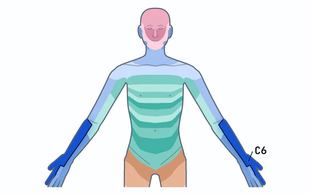
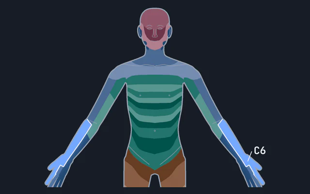
A role says which muscles work; how they make force is the subject of the muscle twitch and tetanus simulator, and why the same muscle is stronger at one joint angle than another is moment arms, which the torque calculator works through. The spinal roots that supply each of these muscles are on the dermatome and myotome map, and the root mode of this page colours the map by them.
Common questions
What muscles do squats work?
In a barbell back squat the quadriceps extend the knee while gluteus maximus and adductor magnus extend the hip, so those three are the prime movers. The hamstrings and soleus assist, and the erector spinae, the abdominal wall and gluteus medius hold the trunk and pelvis steady. Squatting deeper or with a wider stance moves more of the work to the hip extensors (Escamilla 2001; Schoenfeld 2010).
What muscles does the deadlift work?
Gluteus maximus, the hamstrings and adductor magnus extend the hip, the quadriceps drive the bar off the floor, and the erector spinae work isometrically to keep the spine from rounding. The trapezius, latissimus dorsi and the forearm flexors hold the bar close and in the hands. A sumo stance uses more of the vasti and tibialis anterior than a conventional one, which uses more of the gastrocnemius (Escamilla et al. 2002).
What muscles do pull-ups work, and how are chin-ups different?
Latissimus dorsi pulls the upper arm down and back while brachialis and biceps bend the elbow; brachioradialis, teres major, the sternal part of pectoralis major, the lower trapezius, the rhomboids and the posterior deltoid assist. Turning the palms towards you for a chin-up raises biceps and pectoralis major activity, and the overhand pull-up raises lower trapezius activity (Youdas et al. 2010).
What is the difference between a prime mover, a synergist and a stabiliser?
A prime mover produces the lift’s main joint action against the load, a synergist helps it or adds a secondary action, and a stabiliser holds a joint or segment still. In the bench press the pectoralis major, the anterior deltoid and the triceps are prime movers, coracobrachialis assists, and the rotator cuff stabilises the shoulder.
What muscles does the Bulgarian split squat work?
On the front leg the quadriceps, gluteus maximus and adductor magnus are the prime movers. Because one leg carries the load, gluteus medius is more active than in a back squat, holding the pelvis level (McCurdy et al. 2010), with gluteus minimus beneath it. The back leg’s hip flexors are stretched rather than loaded, so they appear as a note rather than a role colour.
Does a muscle lighting up mean that exercise will grow it?
Not necessarily. The colours are roles taken from biomechanics and electromyography studies, and electromyography measures how strongly a muscle is activated, not how much it grows (Vigotsky et al. 2018). Growth depends mostly on weekly volume and how close sets go to failure, and is much the same across a wide range of loads (Schoenfeld et al. 2017), so read the map as what a lift involves, not as a ranking of results.
Why is the 3D model a large download, and will it work on a phone?
The muscles are BodyParts3D’s meshes of a real body, 168 of them, simplified from 10.4 million triangles to 4.9 million so they fit in one scene, and the download, about 18 MB, is printed on the button before anything is fetched. The front and back map shows every lift without the download, and once the 3D model is loaded it stays on your device, with a button to remove it.