Head and Neck Muscles Explorer
Turn 99 muscle meshes of the head and neck in 3D: facial expression, mastication, the hyoid groups and the deep neck, each one separable and named.
Visualiser
Head and neck muscles, 100 structures
Checking this device for a stored copy.
Start here
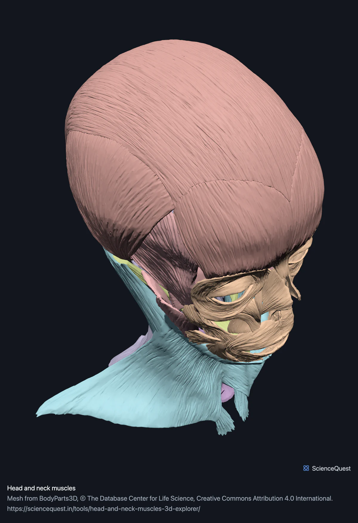
- Hide Scalp and epicranius first One mesh in that band, the aponeurosis over the vault, is a fifth of the model’s triangles on its own. Hiding it exposes the temporalis underneath and makes the model noticeably lighter to turn.
- Click Suprahyoid, then Below The floor of the mouth from underneath, which is the view that makes the mylohyoid sling and the two bellies of the digastric read as a group rather than as separate strips.
- Hide Superficial neck, Suprahyoid and Infrahyoid Strips the neck back to its deep layer: the scalenes, the longus muscles on the front of the spine and the four suboccipital muscles, none of which can be seen from outside.
Muscle, and one aponeurosis. No tendons or fascia beyond the epicranial aponeurosis, no tongue or pharyngeal muscles, no extraocular muscles, and no bone: the skull is a separate model. Muscles of the trunk and limbs have explorers of their own, linked below.
- 100 structures
- 2070k triangles
- 10100 KB, on request
- 0.00241 mm worst error per axis
- 7 scalp and epicranius
- 33 facial expression
- 12 mastication
- 4 superficial neck
- 10 suprahyoid
- 8 infrahyoid
- 26 deep and suboccipital neck
87 muscles in 99 muscle meshes, within 100 meshes. The masseter, lateral pterygoid, orbicularis oculi, digastric and longus colli each arrive in parts, and frontalis and occipitalis are counted separately, as the dataset names them, where Terminologia makes them the two bellies of occipitofrontalis. The remaining mesh is the aponeurosis of the epicranius, the fibrous sheet joining frontalis to occipitalis over the vault, which is not a muscle and is not counted as one.
Where this comes from
Segmented from imaging of one adult male body, not drawn by an artist, so the shapes are measurements. Two consequences worth knowing: it is one person’s anatomy rather than an average, and faint stepping on some surfaces is the resolution of the original scan rather than a rendering fault.
Meshes from BodyParts3D, © The Database Center for Life Science, under Creative Commons Attribution 4.0 International. Names and identifiers are the dataset’s own: Foundational Model of Anatomy identifiers and English labels, as its parts list gives them. Converted meshes are shared under the same licence.
Changes made: Meshes were converted from binary STL to an indexed mesh with 16-bit quantised vertex positions, and surface normals were recomputed from the geometry. No vertex was moved other than by that quantisation, whose worst-case error on any one coordinate is reported on the page.
Latin terms for 68 of the 100 structures come from English Wikipedia anatomy infoboxes, under Creative Commons Attribution-ShareAlike 4.0, joined by Wikidata property P1402, Foundational Model of Anatomy identifier. Only a term a source states for that structure is used, never one constructed from a family name, which is why the rest carry no Latin rather than a guess.
All 100 structures
Every structure the model contains, group by group. Each name opens the viewer with that structure selected, so a link can point at one structure rather than at the whole region.
Scalp and epicranius 7
- aponeurosis of epicranius galea aponeurotica, aponeurosis epicranialis, aponeurosis epicrania other tissue
- left frontalis venter frontalis musculi occipitofrontalis muscle
- left occipitalis venter occipitalis musculi occipitofrontalis muscle
- left temporoparietalis musculus temporoparietalis muscle
- right frontalis venter frontalis musculi occipitofrontalis muscle
- right occipitalis venter occipitalis musculi occipitofrontalis muscle
- right temporoparietalis musculus temporoparietalis muscle
Facial expression 33
- orbital part of left orbicularis oculi muscle
- orbital part of right orbicularis oculi muscle
- palpebral part of left orbicularis oculi muscle
- palpebral part of right orbicularis oculi muscle
- orbicularis oris musculus orbicularis oris muscle
- left zygomaticus major musculus zygomaticus major muscle
- right zygomaticus major musculus zygomaticus major muscle
- left zygomaticus minor musculus zygomaticus minor muscle
- right zygomaticus minor musculus zygomaticus minor muscle
- left levator labii superioris musculus levator labii superioris muscle
- right levator labii superioris musculus levator labii superioris muscle
- left levator labii superioris alaeque nasi m. levator nasolabialis, musculus levator labii superioris alaeque nasi muscle
- right levator labii superioris alaeque nasi m. levator nasolabialis, musculus levator labii superioris alaeque nasi muscle
- left levator anguli oris musculus levator anguli oris, musculus caninus muscle
- right levator anguli oris musculus levator anguli oris, musculus caninus muscle
- left depressor labii inferioris musculus depressor labii inferioris muscle
- right depressor labii inferioris musculus depressor labii inferioris muscle
- left depressor anguli oris musculus depressor anguli oris muscle
- right depressor anguli oris musculus depressor anguli oris muscle
- left mentalis musculus mentalis muscle
- right mentalis musculus mentalis muscle
- left risorius muscle
- right risorius muscle
- left buccinator muscle
- right buccinator muscle
- left procerus musculus procerus, pyramidalis nasi, depressor glabellae muscle
- right procerus musculus procerus, pyramidalis nasi, depressor glabellae muscle
- left nasalis musculus nasalis muscle
- right nasalis musculus nasalis muscle
- left depressor septi nasi musculus depressor septi nasi muscle
- right depressor septi nasi musculus depressor septi nasi muscle
- left corrugator supercilii musculus corrugator supercilii muscle
- right corrugator supercilii musculus corrugator supercilii muscle
Mastication 12
- deep part of left masseter muscle
- deep part of right masseter muscle
- superficial part of left masseter muscle
- superficial part of right masseter muscle
- left temporalis musculus temporalis muscle
- right temporalis musculus temporalis muscle
- lower head of left lateral pterygoid muscle
- lower head of right lateral pterygoid muscle
- upper head of left lateral pterygoid muscle
- upper head of right lateral pterygoid muscle
- left medial pterygoid musculus pterygoideus medialis, musculus pterygoideus internus muscle
- right medial pterygoid musculus pterygoideus medialis, musculus pterygoideus internus muscle
Superficial neck 4
- left platysma platysma muscle
- right platysma platysma muscle
- left sternocleidomastoid musculus sternocleidomastoideus muscle
- right sternocleidomastoid musculus sternocleidomastoideus muscle
Suprahyoid 10
- anterior belly of left digastric muscle
- anterior belly of right digastric muscle
- posterior belly of left digastric muscle
- posterior belly of right digastric muscle
- left mylohyoid musculus mylohyoideus muscle
- right mylohyoid musculus mylohyoideus muscle
- left geniohyoid musculus geniohyoideus muscle
- right geniohyoid musculus geniohyoideus muscle
- left stylohyoid musculus stylohyoideus muscle
- right stylohyoid musculus stylohyoideus muscle
Infrahyoid 8
- left sternohyoid musculus sternohyoideus muscle
- right sternohyoid musculus sternohyoideus muscle
- left sternothyroid musculus sternothyroideus muscle
- right sternothyroid musculus sternothyroideus muscle
- left thyrohyoid musculus thyrohyoideus muscle
- right thyrohyoid musculus thyrohyoideus muscle
- left omohyoid musculus omohyoideus muscle
- right omohyoid musculus omohyoideus muscle
Deep and suboccipital neck 26
- left scalenus anterior muscle
- right scalenus anterior muscle
- left scalenus medius muscle
- right scalenus medius muscle
- left scalenus posterior muscle
- right scalenus posterior muscle
- inferior oblique part of left longus colli muscle
- inferior oblique part of right longus colli muscle
- superior oblique part of left longus colli muscle
- superior oblique part of right longus colli muscle
- vertical intermediate part of left longus colli muscle
- vertical intermediate part of right longus colli muscle
- left longus capitis musculus longus capitis muscle
- right longus capitis musculus longus capitis muscle
- left rectus capitis anterior musculus rectus capitis anterior muscle
- right rectus capitis anterior musculus rectus capitis anterior muscle
- left rectus capitis lateralis musculus rectus capitis lateralis muscle
- right rectus capitis lateralis musculus rectus capitis lateralis muscle
- left rectus capitis posterior major musculus rectus capitis posterior major muscle
- right rectus capitis posterior major musculus rectus capitis posterior major muscle
- left rectus capitis posterior minor musculus rectus capitis posterior minor muscle
- right rectus capitis posterior minor musculus rectus capitis posterior minor muscle
- left obliquus capitis superior musculus obliquus capitis superior muscle
- right obliquus capitis superior musculus obliquus capitis superior muscle
- left obliquus capitis inferior musculus obliquus capitis inferior muscle
- right obliquus capitis inferior musculus obliquus capitis inferior muscle
Citing this tool
Last updated . Add the date you accessed it as well, which a citation of a page that can change asks for. If a specific view matters, cite the page address as it stands in your browser instead: it records what is selected and shown.
Why the head and neck, and not the whole body
Because every muscle at once does not fit in one scene. The muscle models on this site come to 18.3 million triangles between them, more than twice what the viewer will load without asking first, so the musculature ships one region at a time. This is the head and neck: 99 muscle meshes, 10 megabytes and 2.07 million triangles.
The other regions have explorers of their own: the upper limb, lower limb, chest wall, abdominal wall and pelvic floor and back. The full body explorer can lay any of them over the skeleton as a layer.
The bands are functional, and the completeness is checked
Every other region here derives its part list from the dataset’s part-of hierarchy, which is what stops a part being lost by hand. That cannot produce these bands: under musculature of head the hierarchy goes straight from the region to individual named muscles, with no node for the muscles of facial expression or the muscles of mastication. Those are functional groupings that anatomists use and the data does not record.
So each band names the muscles it holds, and the generator then asserts that the union of all seven bands is exactly the set of leaves under musculature of head and musculature of neck, in both directions. A muscle missing from every band fails, and an invented one fails too. The grouping is a judgement; the completeness is not.
One structure here is not a muscle
The aponeurosis of the epicranius, the galea, is the fibrous sheet that joins the frontalis in front to the occipitalis behind and lets them move the scalp as one. It is tendon rather than muscle, so it is tagged as other tissue and left out of the muscle count, and it sits in the scalp band with the bellies it connects.
Muscles and bones are in the same coordinate frame
This model is the muscles alone, so a muscle and its bony attachment are not in one view here. The head and neck explorer puts them in one: it loads these same meshes together with the skull, the cervical spine, the teeth and the brain. Nothing is fitted to make that work. Every model on this site comes from the same segmentation of the same body in one shared coordinate frame, so the temporalis lands in the temporal fossa the same millimetres from the bone as in a body, rather than as two drawings at two arbitrary scales.
Common questions
Why only the head and neck, and not every muscle in the body?
Because every muscle at once does not fit in one scene. The six muscle models on this site together come to more than twice the triangles the viewer will load without asking first, so the musculature ships one region at a time, and this is the head and neck: 99 muscle meshes, with the download size printed on the load button. The upper limb, lower limb, chest wall, abdominal wall and back each have an explorer of their own, and the full body explorer can lay any of them over the skeleton as a layer.
Is every head and neck muscle really here?
Every one the dataset publishes for the head and neck is, and that is checked rather than claimed. The bands are declared as groups of named muscles, and the build asserts that their union is exactly the set of meshes the dataset files under musculature of head and musculature of neck, in both directions. A muscle left out of a band fails the build instead of quietly vanishing from the model, which is the same check the skeleton uses. Some muscles of the region are still not here: the dataset publishes no tongue, pharyngeal, laryngeal, palatal, extraocular or auricular muscles, and the trapezius, levator scapulae, splenius capitis and semispinalis capitis at the back of the neck are in the back muscles explorer. One structure in it is not a muscle: the aponeurosis of the epicranius is the fibrous sheet joining the frontalis and occipitalis over the vault, so it is tagged as other tissue and not counted among the muscles.
Why is the scalp band so much bigger than the rest?
One mesh in it, the aponeurosis, is a fifth of the whole model’s triangles on its own. Thin broad sheets are the worst case for a triangle budget, exactly as the ribs and scapulae are in the skeleton: a surface that encloses almost no volume has nothing to amortise its own area against. Hiding the scalp band is therefore also the quickest way to make the model lighter to turn on a slow device.
Can I see what a muscle attaches to?
Not on this page, which is muscle only, but the head and neck explorer shows it. The dataset publishes muscles and bones as separate meshes in one shared coordinate frame, so they sit in the right places relative to each other, and that page loads these muscles in one scene with the skull, the cervical spine, the teeth and the brain: a temporalis there sits in its temporal fossa as it does in a body.