Lungs and Bronchial Tree 3D Explorer
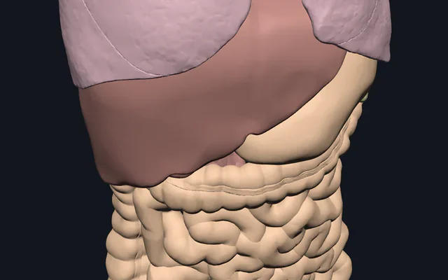
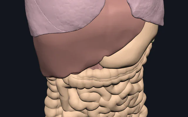
Turn real 3D lungs inside the rib cage: the five lobes and their fissures, the trachea and bronchial tree, and the pulmonary arteries and veins.
Visualiser
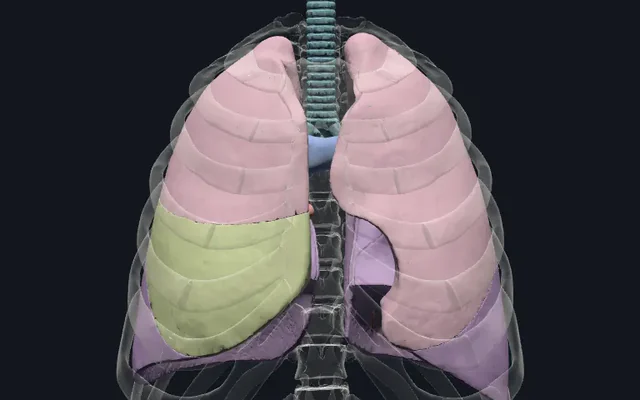
Lungs and bronchial tree, 9 structures
2.2 MB, downloaded only when you press Start exploring and kept on this device afterwards.
Start here
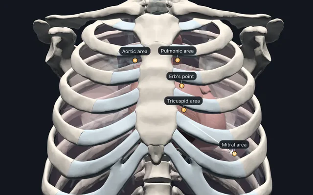
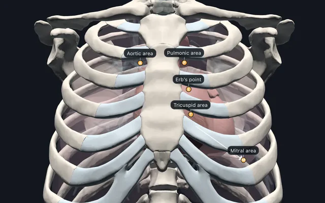
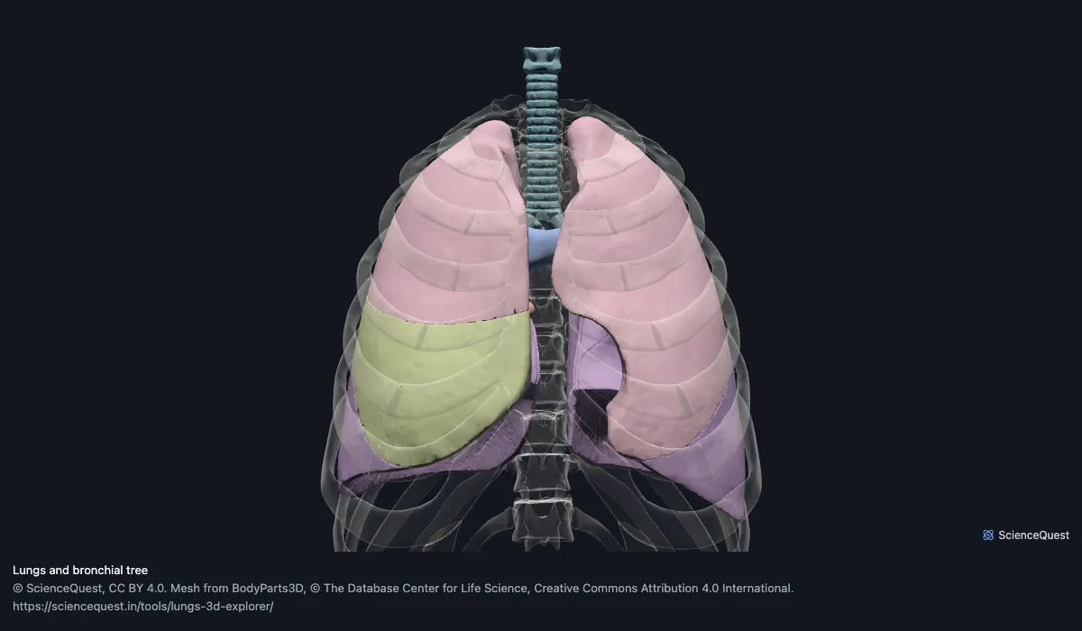
- Turn to the front and right The ribs, sternum and spine as glass from three-quarters: the right lung’s three lobes and both of its fissures inside the cage.
- Hide the bony thorax The five lobes, coloured by level, so every fissure is a gap between two colours.
- Show the right lung’s fissures From the right: the horizontal fissure above the middle lobe and the oblique fissure behind it.
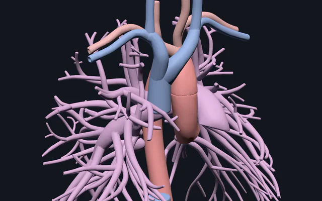
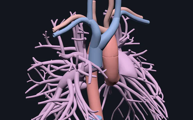
- Hide the lobes The trachea, the bronchial tree and both pulmonary vessel trees, branching into each lung from the hilum.
Lungs, airway and pulmonary vessels, in the bony thorax. The bronchial tree and each pulmonary vessel tree are one mesh each in BodyParts3D 3.0, the release these meshes come from, so lobar and segmental bronchi are not separate and cannot be picked one by one. There are no fissure meshes either: each fissure is the gap between two lobe meshes. No pleura, no heart, no diaphragm and no bronchial arteries. The heart the pulmonary vessels run to is in the heart 3D explorer, and 14 listening sites over the lung fields are in the auscultation points explorer.
9 structures: 2 upper lobes, 1 middle lobe, 2 lower lobes, the trachea, the bronchus, the pulmonary artery and the pulmonary vein.
The 55 meshes of the bony thorax load with them for context and are not counted: 12 thoracic vertebrae, 3 parts of the sternum, 24 ribs, 16 costal cartilages. All 64 are listed, nameable and selectable. Simplified from 1,513,552 triangles to 558,716, and no original vertex lies more than 0.566 mm from the simplified surface, measured mesh by mesh.
Where this comes from
Segmented from MRI of one adult male volunteer, then refined by medical illustrators, who filled in missing detail and sharpened blurred outlines from textbooks and atlases (Mitsuhashi et al., Nucleic Acids Research, 2009). Two consequences worth knowing: it is one person’s anatomy rather than an average, and faint stepping on some surfaces is the resolution of the original scan rather than a rendering fault.
Meshes from BodyParts3D, © The Database Center for Life Science, under Creative Commons Attribution 4.0 International. Names and identifiers are the dataset’s own: Foundational Model of Anatomy identifiers and English labels, as its parts list gives them. Converted meshes are shared under the same licence.
Changes made: Meshes were converted from binary STL to an indexed mesh with 16-bit quantised vertex positions, and surface normals were recomputed from the geometry. No vertex was moved other than by that quantisation, whose worst-case error on any one coordinate is reported on the page. On this page it is at most 0.0156 mm per axis. This model was then made from a selection of those converted meshes: each was simplified by edge collapse that keeps only original vertices, at the largest tolerance whose measured distance from every original vertex to the simplified surface stays within 0.7 mm, or left whole where none did, and its positions were quantised again to 16 bits over this model’s own bounds. Each model’s file is then compressed without loss, by meshoptimizer’s vertex and index codecs and gzip, and restored exactly before it is drawn. The file holds 558,716 triangles in 2,239 kB, downloaded only when you press Start exploring.
Latin terms for 5 of the 64 structures come from English Wikipedia anatomy infoboxes, under Creative Commons Attribution-ShareAlike 4.0, joined by Wikidata property P1402, Foundational Model of Anatomy identifier. Only a term a source states for that structure is used, never one constructed from a family name, which is why the rest carry no Latin rather than a guess. A few are corrected to the Terminologia Anatomica form where Wikipedia gives an older one, and where several forms are listed, some may still be older synonyms rather than the Terminologia term.
Lobes, fissures and airways: the sources and this model
What the anatomy texts say beside what this model shows, and where to look. Each name selects that structure in the 3D lungs above; every figure in the last column is measured on the model’s own meshes.
| Feature | What the sources say | In this model |
|---|---|---|
| Right lung | Three lobes, upper, middle and lower, parted by two fissures. The middle lobe is the smallest and wedge-shaped. (Gray 1918, The Lungs; StatPearls, Lungs) | Three lobe meshes; no fissure meshes, so each fissure is the gap between two lobes. upper lobe of right lung, middle lobe of lung, lower lobe of right lung |
| Left lung | Two lobes, upper and lower, parted by one fissure; the lower is the larger. The anterior border has the cardiac notch. (Gray 1918, The Lungs; StatPearls, Lungs) | Two lobe meshes; the notch is on the front of the upper lobe. upper lobe of left lung, lower lobe of left lung |
| Oblique fissure | Both lungs. It separates the lower lobe from the lobes above and in front; on the left it crosses the posterior border about 6 cm below the apex. (Gray 1918, The Lungs; StatPearls, Lungs) | The gap between each lower lobe and the lobes above and in front of it. lower lobe of right lung, lower lobe of left lung |
| Horizontal fissure | Right lung only. It separates the upper from the middle lobe and meets the anterior border level with the sternal end of the fourth costal cartilage. (Gray 1918, The Lungs) | Here the front of the middle lobe reaches 2 mm above the sternal end of the right fourth costal cartilage. middle lobe of lung, right fourth costal cartilage |
| Height | The right lung is broader than the left but about 2.5 cm shorter, because the diaphragm rises higher on the right. (Gray 1918, The Lungs) | Here the right base sits 14 mm higher than the left, taking the base as the median lowest point over a 5 mm grid seen from above, where the lung is at least 120 mm deep. Top to bottom the two are 254 mm and 259 mm, close because that height runs to the one lowest point of each base, and those two points are within 3 mm of each other. |
| Trachea | From the larynx, level with the sixth cervical vertebra, to its division into the two main bronchi: at the upper border of the fifth thoracic vertebra in Gray; at the sternal angle, level with the fourth thoracic vertebra, in StatPearls. About 11 cm long. (Gray 1918, The Trachea and Bronchi; StatPearls, Bronchial) | This trachea mesh is 118 mm tall and ends 3 mm below the top of the body of the fifth thoracic vertebra, level with the sternal angle. trachea |
| Main bronchi | The right is wider, shorter (about 2.5 cm) and more vertical, so an inhaled foreign body more often enters it; the left is narrower, nearly 5 cm long, and passes beneath the aortic arch. (Gray 1918, The Trachea and Bronchi; StatPearls, Bronchial) | Part of the one bronchial tree mesh, both sides. bronchus |
| Lobar and segmental bronchi | One lobar bronchus per lobe, three right and two left. Each segmental bronchus supplies a bronchopulmonary segment: 10 in the right lung, and 8 to 9 (StatPearls, Lungs) or 8 to 10 (StatPearls, Bronchial) in the left. (StatPearls, Bronchial; StatPearls, Lungs) | Not separate meshes: the branches are all inside the one bronchial tree mesh. bronchus |
| Root of the lung | Front to back, the upper pulmonary vein, the pulmonary artery, then the bronchus. Top to bottom, on the right the upper lobe (eparterial) bronchus, the artery, the rest of the bronchus, the veins; on the left the artery, the bronchus, the veins. (Gray 1918, The Lungs; Gray 1918, The Trachea and Bronchi; StatPearls, Lungs) | Hide the lobes to see the three trees enter each lung. pulmonary vein, pulmonary artery, bronchus |
| Pulmonary vessels | The pulmonary artery leaves the right ventricle and divides into right and left pulmonary arteries, whose branches follow the bronchi. Four pulmonary veins join the left atrium. (StatPearls, Lungs; Gray 1918, The Lungs) | One mesh for the arterial tree and one for the venous tree; the heart is not in this model. pulmonary artery, pulmonary vein |
Sources, for this table and the common questions below: Gray H. Anatomy of the Human Body. 20th ed., revised by Lewis WH. Philadelphia: Lea & Febiger; 1918, XI Splanchnology, 1e, The Lungs (Bartleby.com edition, archived copy); Gray H. Anatomy of the Human Body. 20th ed., revised by Lewis WH. Philadelphia: Lea & Febiger; 1918, XI Splanchnology, 1b, The Trachea and Bronchi (Bartleby.com edition, archived copy); Chaudhry R, Omole AE, Bordoni B. Anatomy, Thorax, Lungs. StatPearls. Treasure Island (FL): StatPearls Publishing (updated ); Amador C, Weber C, Varacallo MA. Anatomy, Thorax, Bronchial. StatPearls. Treasure Island (FL): StatPearls Publishing (updated ).
All 64 structures
Every structure the model contains, group by group. Of these, 9 are structures of this region. Each name opens the viewer with that structure selected, so a link can point at one structure rather than at the whole region.
Upper lobes 2
- upper lobe of right lung page organ
- upper lobe of left lung page organ
Middle lobe 1
- middle lobe of lung page organ
Lower lobes 2
- lower lobe of right lung page organ
- lower lobe of left lung page organ
Pulmonary artery 1
- pulmonary artery page arteria pulmonalis blood vessel
Pulmonary vein 1
- pulmonary vein page venae pulmonales blood vessel
Thoracic vertebrae 12
- first thoracic vertebra page
- second thoracic vertebra page
- third thoracic vertebra page
- fourth thoracic vertebra page
- fifth thoracic vertebra page
- sixth thoracic vertebra page
- seventh thoracic vertebra page
- eighth thoracic vertebra page
- ninth thoracic vertebra page
- tenth thoracic vertebra page
- eleventh thoracic vertebra page
- twelfth thoracic vertebra page
Sternum 3
- manubrium page
- body of sternum page
- xiphoid process page processus xiphoideus
Ribs 24
- right first rib page
- left first rib page
- right second rib page
- left second rib page
- right third rib page
- left third rib page
- right fourth rib page
- left fourth rib page
- right fifth rib page
- left fifth rib page
- right sixth rib page
- left sixth rib page
- right seventh rib page
- left seventh rib page
- right eighth rib page
- left eighth rib page
- right ninth rib page
- left ninth rib page
- right tenth rib page
- left tenth rib page
- right eleventh rib page
- left eleventh rib page
- right twelfth rib page
- left twelfth rib page
Costal cartilage 16
- right first costal cartilage page cartilage
- left first costal cartilage page cartilage
- right second costal cartilage page cartilage
- left second costal cartilage page cartilage
- right third costal cartilage page cartilage
- left third costal cartilage page cartilage
- right fourth costal cartilage page cartilage
- left fourth costal cartilage page cartilage
- right fifth costal cartilage page cartilage
- left fifth costal cartilage page cartilage
- right sixth costal cartilage page cartilage
- left sixth costal cartilage page cartilage
- right seventh costal cartilage page cartilage
- left seventh costal cartilage page cartilage
- right costal cartilage page cartilage
- left costal cartilage page cartilage
Cite this tool
Citing this tool
Last updated . Add the date you accessed it as well, which a citation of a page that can change asks for. If a specific view matters, cite the page address as it stands in your browser instead: it records what is selected and shown.
On the syllabus: MCAT, 3B: Structure and integrative functions of the main organ systems.
How many lobes does each lung have?
Three on the right, upper, middle and lower, and two on the left, upper and lower (Gray 1918; StatPearls, Lungs). Here the two upper lobes share a colour, the middle lobe has its own and the two lower lobes share a third, so the lobes on either side of any fissure always differ. The oblique fissure, in both lungs, is the boundary of the lower lobe; the horizontal fissure, on the right only, runs between the upper and middle lobes (Gray 1918).
Gray places the front of the horizontal fissure level with the sternal end of the fourth costal cartilage. On this model the front of the middle lobe reaches 2 mm above that point. The trachea mesh ends 3 mm below the top of the body of the fifth thoracic vertebra, level with the sternal angle; Gray puts the division of the trachea at the upper border of the fifth thoracic vertebra, and StatPearls, Bronchial at the sternal angle, level with the fourth, so this model agrees with both.
One mesh for the whole bronchial tree
The bronchial tree and each pulmonary vessel tree are one mesh each in BodyParts3D 3.0; lobar and segmental bronchi are not separate. Picking the bronchus selects both main bronchi and every branch the dataset segmented. StatPearls, Bronchial counts one lobar bronchus per lobe, and a segmental bronchus for each bronchopulmonary segment, 10 on the right and 8 to 10 on the left (8 to 9 in StatPearls, Lungs); those branches are inside the one mesh, so to follow them, hide the lobes, glass the vessels and turn the model. BodyParts3D 4.0 does segment the main and segmental bronchi, but it has no lobe surfaces and is in a different frame, so its meshes cannot be combined with these.
At the hilum
Each lung’s root carries its bronchus, pulmonary artery and pulmonary veins. From front to back the order is the upper pulmonary vein, the artery, then the bronchus (Gray 1918; StatPearls, Lungs). From above down it differs by side: on the right the upper lobe bronchus comes off above the artery, which is why Gray calls it eparterial, while on the left the artery lies above the bronchus. The arteries are blue here and the veins red because the pulmonary artery carries deoxygenated blood from the right ventricle to the lungs, and the four pulmonary veins return oxygenated blood to the left atrium (StatPearls, Lungs; Gray 1918).
What the model leaves out
- Separate bronchi and vessel branches. One mesh each for the bronchial tree, the pulmonary arteries and the pulmonary veins.
- Fissures as surfaces. The fissures are the gaps between the lobe meshes.
- Breathing. The lungs are one static shape from one scan.
- One body. The meshes are one adult man’s, so every size here is his.
Common questions
How many lobes does each lung have?
The right lung has three, upper, middle and lower, and the left has two, upper and lower (Gray 1918; StatPearls, Anatomy, Thorax, Lungs). The left lung’s lower lobe is the larger of its two, and the right middle lobe is the smallest lobe of the right lung (Gray 1918).
What do the oblique and horizontal fissures separate?
The oblique fissure, in both lungs, separates the lower lobe from the lobes above and in front of it. The horizontal fissure is in the right lung only; it separates the upper from the middle lobe and meets the front of the lung level with the sternal end of the fourth costal cartilage (Gray 1918). Measured on this model, the front of the middle lobe reaches 2 mm above that point.
Why do inhaled objects usually go into the right bronchus?
The right main bronchus is wider, shorter and more vertical than the left, so it continues the line of the trachea more directly (Gray 1918; StatPearls, Anatomy, Thorax, Bronchial).
Where does the trachea divide?
Gray (1918) puts the division at the upper border of the fifth thoracic vertebra; StatPearls, Anatomy, Thorax, Bronchial puts it at the sternal angle, level with the fourth thoracic vertebra. Measured on this model, the trachea mesh ends 3 mm below the top of the body of the fifth thoracic vertebra, level with the sternal angle, so it agrees with both.
Are the segmental bronchi in the model?
Not as separate structures. In BodyParts3D 3.0 the whole bronchial tree is one mesh, and so is each pulmonary vessel tree. StatPearls, Anatomy, Thorax, Bronchial gives 10 bronchopulmonary segments in the right lung and 8 to 10 in the left (8 to 9 in StatPearls, Anatomy, Thorax, Lungs), each supplied by its own segmental bronchus.