Trachea
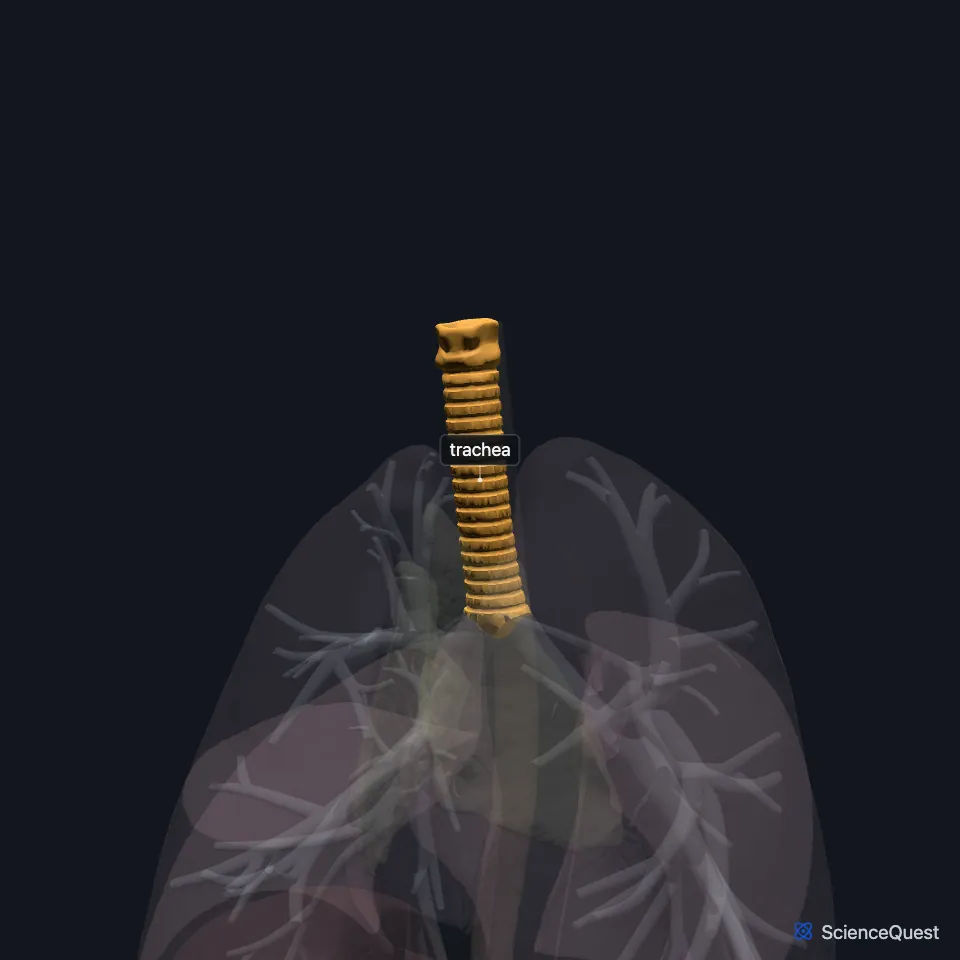
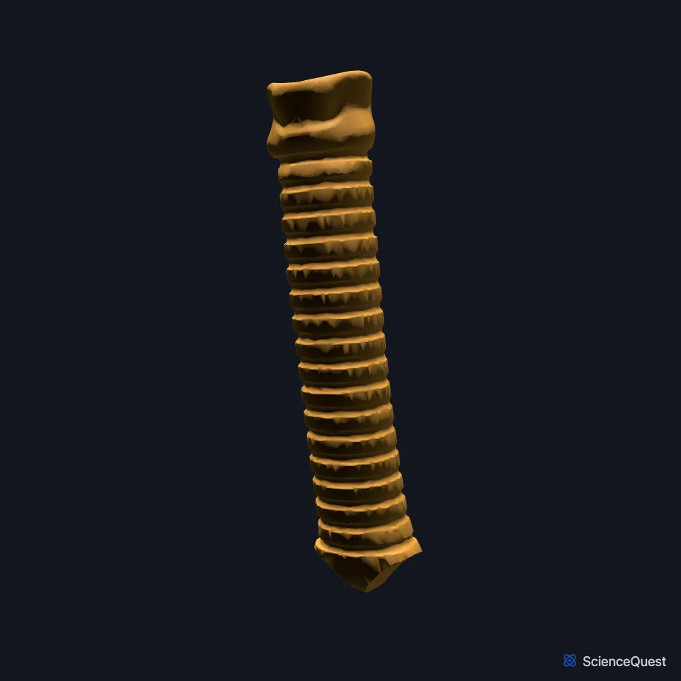
The trachea, or windpipe, is the airway that runs from the larynx toward the lungs. Stacked, C-shaped pieces of hyaline cartilage hold it open. (OpenStax A&P 2e, 22.1; StatPearls, Trachea and Bronchial)
Where is the trachea?
It begins just below the larynx, level with the cricoid cartilage and the sixth cervical vertebra. It ends at the carina, level with the fourth to fifth thoracic vertebrae, where it splits into the two main bronchi. (StatPearls, Trachea)
An adult trachea is about 10 to 13 cm long. In infants it measures about 5 to 6 cm. (StatPearls, Trachea)
Its upper, cervical part is about 2 to 4 cm long and gives way to the thoracic part at about the sixth tracheal ring. (StatPearls, Trachea)
What is the trachea made of?
Its wall has four layers: a mucosa of pseudostratified ciliated columnar epithelium, a submucosa of connective tissue, a musculocartilaginous layer, and an outer adventitia that anchors it to the tissues around it. (StatPearls, Trachea)
The 16 to 20 cartilage rings cover the front and sides and stop it collapsing as you breathe in. At the back, the trachealis muscle and elastic tissue form a fibroelastic membrane that lets the trachea stretch a little with each breath. (StatPearls, Trachea; OpenStax A&P 2e, 22.1)
Goblet cells in the lining release mucus that traps particles. Cilia then sweep the mucus up toward the oropharynx, a process called mucociliary clearance. (StatPearls, Trachea)
What does the trachea do?
It is the main passage for air between the larynx and the lungs, on every breath in and out. (StatPearls, Trachea)
Contracting the trachealis narrows the trachea and raises the pressure inside it, which helps force air out when you cough. When the muscle relaxes, the soft back wall of the trachea can move, and the esophagus behind it has room to expand as you swallow. (StatPearls, Trachea; OpenStax A&P 2e, 22.1)
As you swallow, breathing pauses and the vocal folds close. A cough helps clear food that gets into the airway anyway. (OpenStax A&P 2e, 23.3; StatPearls, Physiology, Swallowing and Trachea)
What lies around the trachea?
The esophagus lies directly behind it, and the recurrent laryngeal nerves climb in the grooves between the two. (StatPearls, Trachea; OpenStax A&P 2e, 22.1)
In front of it lie the inferior thyroid veins and the left brachiocephalic vein. Beside it, near the sixth ring, lie the lobes of the thyroid gland and the carotid sheaths, which carry the common carotid artery, the internal jugular vein and the vagus nerve. (StatPearls, Trachea)
Its blood comes mainly from the inferior thyroid and bronchial arteries. The thoracic part is fed by bronchial arteries from the thoracic aorta, level with the fifth and sixth thoracic vertebrae. (StatPearls, Trachea)
The vagus nerve and its recurrent laryngeal branches bring parasympathetic fibers, which contract the airway muscle and increase gland secretion. Sympathetic fibers from the cervical and upper thoracic ganglia relax it. (StatPearls, Trachea)
What can go wrong as the trachea forms?
In the embryo a septum splits one foregut tube into the trachea in front and the esophagus behind. If it fails to form or fuse, the result is one of a group of defects, among them esophageal atresia and tracheoesophageal fistula, that together occur in about 1 in 3,000 live births. (StatPearls, Trachea)
Where the sources differ
The sources differ on what mainly keeps food out of the airway. StatPearls names closure of the vocal folds as the main guard and says the epiglottis is not directly involved in airway protection, while OpenStax credits the epiglottis folding down over the larynx. (StatPearls, Physiology, Swallowing; OpenStax A&P 2e, 23.3)
The two StatPearls articles place the carina a little differently: the trachea article at the fourth to fifth thoracic vertebrae, the bronchi article at the sternal angle, level with the fourth thoracic vertebra. (StatPearls, Trachea and Bronchial)
| Feature | Detail and source |
|---|---|
| Begins | Below the larynx, at the cricoid cartilage and C6 (StatPearls, Trachea) |
| Ends | At the carina, T4 to T5, as the two main bronchi (StatPearls, Trachea) |
| Length | About 10 to 13 cm in adults (StatPearls, Trachea) |
| Cartilage | 16 to 20 C-shaped rings of hyaline cartilage (OpenStax A&P 2e, 22.1; StatPearls, Trachea) |
| Arteries | Inferior thyroid and bronchial arteries (StatPearls, Trachea) |
| Nerves | Vagus and recurrent laryngeal nerves; cervical and upper thoracic sympathetic ganglia (StatPearls, Trachea) |
Touches in this model
Measured on the BodyParts3D meshes: surfaces within 2 mm of the trachea, nearest first.
- Bronchus in contact
- Esophagus in contact
- Arch of aorta in contact
- Left thyrohyoid 0.8 mm away
- Right thyrohyoid 0.8 mm away
- Left sternothyroid 1.0 mm away
- Right sternothyroid 1.0 mm away
- Upper lobe of right lung 1.4 mm away
- Skin 1.5 mm away
- Vertical intermediate part of right longus colli 2.0 mm away
Sources
- OpenStax Anatomy and Physiology 2e, section 22.1, Organs and Structures of the Respiratory System. CC BY-NC-SA 4.0. Read 2026-10-25.
- StatPearls, Anatomy, Head and Neck, Trachea. Hayley Derricott; Jonathan Jimenez; James La Rose. Last update June 8, 2026. CC BY-NC-ND 4.0. Read 2026-10-25.
- StatPearls, Anatomy, Thorax, Bronchial. Christina Amador; Carly Weber; Matthew A. Varacallo. Last update August 8, 2023. CC BY-NC-ND 4.0. Read 2026-10-25.
- OpenStax Anatomy and Physiology 2e, section 23.3, The Mouth, Pharynx, and Esophagus. CC BY-NC-SA 4.0. Read 2026-10-25.
- StatPearls, Physiology, Swallowing. Kush Panara; Edris Ramezanpour Ahangar; Devang Padalia. Last update July 24, 2023. CC BY-NC-ND 4.0. Read 2026-10-25.
The values are facts and free to use; this page’s selection and presentation are © 2026 ScienceQuest.