Esophagus
The esophagus is a muscular tube, about 25 cm (10 in) long in adults, that connects the pharynx to the stomach. (OpenStax A&P 2e, 23.3; Gray’s Anatomy (1918), The Esophagus; StatPearls, Physiology, Lower Esophageal Sphincter)
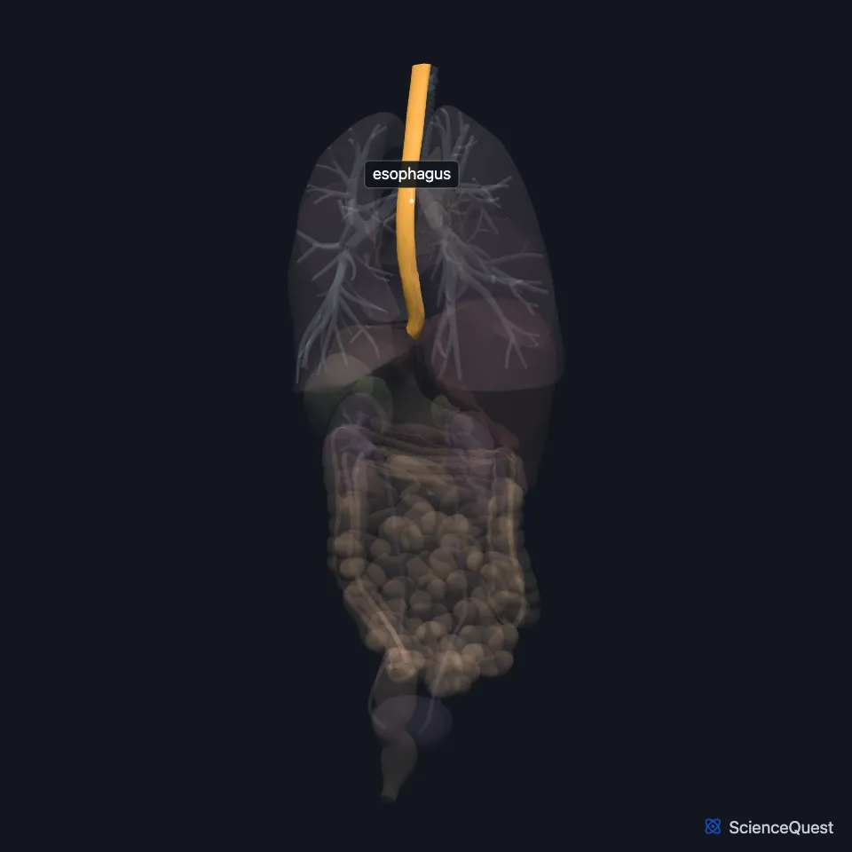
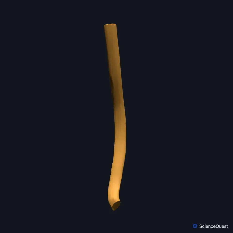
Where is the esophagus?
It runs behind the trachea and the heart and in front of the spine, then crosses the diaphragm to reach the stomach. (StatPearls, Esophagus)
It has cervical, thoracic and abdominal parts. The cervical part runs from the cricopharyngeus to the suprasternal notch, the thoracic part from there to the diaphragm, and the abdominal part from the diaphragm to the cardia of the stomach. (StatPearls, Esophagus)
Level with the discs between the fourth and fifth thoracic vertebrae it passes the arch of the aorta and enters the posterior mediastinum. It goes through the diaphragm at the esophageal hiatus, at about the tenth thoracic vertebra. (StatPearls, Esophagus)
What is the esophagus made of?
Its lining is stratified squamous epithelium, like the skin’s but non-keratinized. Small mucous glands lie in the wall beneath it. (OpenStax A&P 2e, 23.3; StatPearls, Physiology, Esophagus)
Its muscle has an outer longitudinal layer and a thicker inner circular layer, which does the work of peristalsis. At the lower end the circular fibers thicken and overlap to form the lower esophageal sphincter. (StatPearls, Esophagus)
The outermost layer is adventitia, a fibrous coat that binds the esophagus to the structures around it. (OpenStax A&P 2e, 23.3; StatPearls, Physiology, Esophagus; Gray’s Anatomy (1918), The Esophagus)
What does the esophagus do?
The upper esophageal sphincter controls entry from the pharynx. A peristaltic wave that starts in the pharynx carries on down the esophagus and pushes each mouthful toward the stomach. (OpenStax A&P 2e, 23.3; StatPearls, Esophagus)
If the first wave fails to clear the bolus, the stretched esophagus sets off secondary peristalsis, which keeps going until the bolus reaches the stomach. (StatPearls, Esophagus)
The lower esophageal sphincter stays contracted to keep stomach contents out and relaxes after you swallow to let food through. If it does not close fully, stomach contents can reflux and cause heartburn or gastroesophageal reflux disease (GERD). (OpenStax A&P 2e, 23.3; StatPearls, Esophagus and Physiology, Lower Esophageal Sphincter)
What supplies the esophagus, and how does it form?
Each part has its own arteries: the inferior thyroid artery for the cervical part, esophageal branches of the thoracic aorta for the thoracic part, and the left gastric and left phrenic arteries for the abdominal part. (StatPearls, Esophagus)
Its veins drain in segments too: to the brachiocephalic veins from the neck, to the azygos and hemiazygos veins in the chest, and through the left gastric vein to the portal system at the lower end. (StatPearls, Esophagus)
The vagus nerve drives its muscle and glands. Sympathetic fibers from the thoracic and cervical trunks keep up the tone of both sphincters and generally slow peristalsis. (StatPearls, Esophagus)
The esophagus and the trachea start as one foregut tube in the embryo. A septum divides it into the trachea in front and the esophagus behind. (StatPearls, Trachea)
Where the sources differ
The sources split its muscle differently. OpenStax gives skeletal muscle in the upper third, a mix in the middle third and smooth muscle in the lower third; StatPearls gives mainly skeletal muscle in the upper third and smooth muscle in the lower two thirds. (OpenStax A&P 2e, 23.3; StatPearls, Esophagus)
They also give slightly different lengths: OpenStax about 25.4 cm (10 in), StatPearls 9 to 10 in in adults. (OpenStax A&P 2e, 23.3; StatPearls, Esophagus)
| Feature | Detail and source |
|---|---|
| Parts | Cervical, thoracic, abdominal (StatPearls, Esophagus) |
| Crosses the diaphragm | At the esophageal hiatus, about T10 (StatPearls, Esophagus) |
| Sphincters | Upper esophageal sphincter at the pharynx; lower esophageal sphincter at the stomach (OpenStax A&P 2e, 23.3; StatPearls, Physiology, Esophagus) |
| Arteries | Inferior thyroid, esophageal branches of the thoracic aorta, left gastric, left phrenic (StatPearls, Esophagus) |
| Nerves | Vagus nerve; thoracic and cervical sympathetic trunks (StatPearls, Esophagus) |
Touches in this model
Measured on the BodyParts3D meshes: surfaces within 2 mm of the esophagus, nearest first, 12 of 33.
- Lower lobe of right lung in contact
- Stomach in contact
- Trachea in contact
- Descending aorta in contact
- Diaphragm in contact
- Fifth cervical vertebra in contact
- Fifth thoracic vertebra in contact
- First thoracic vertebra in contact
- Inferior oblique part of left longus colli in contact
- Intervertebral disk of fifth cervical vertebra in contact
- Intervertebral disk of fifth thoracic vertebra in contact
- Intervertebral disk of seventh cervical vertebra in contact
Sources
- OpenStax Anatomy and Physiology 2e, section 23.3, The Mouth, Pharynx, and Esophagus. CC BY-NC-SA 4.0. Read 2026-10-25.
- Gray’s Anatomy of the Human Body, 20th US edition (1918), Splanchnology, The Esophagus, pages 1144 to 1146. Henry Gray; edited by Warren H. Lewis. Public domain. Read 2026-10-25.
- StatPearls, Physiology, Lower Esophageal Sphincter. Ryan D. Rosen; Ryan Winters. Last update March 17, 2023. CC BY-NC-ND 4.0. Read 2026-10-25.
- StatPearls, Anatomy, Thorax, Esophagus. Adekunle E. Omole; Bruno Bordoni. Last update January 31, 2026. CC BY-NC-ND 4.0. Read 2026-10-25.
- StatPearls, Physiology, Esophagus. Suhaib A. Bajwa; Fadi Toro; Anup Kasi. Last update May 1, 2023. CC BY-NC-ND 4.0. Read 2026-10-25.
- StatPearls, Anatomy, Head and Neck, Trachea. Hayley Derricott; Jonathan Jimenez; James La Rose. Last update June 8, 2026. CC BY-NC-ND 4.0. Read 2026-10-25.
The values are facts and free to use; this page’s selection and presentation are © 2026 ScienceQuest.