Diaphragm
The diaphragm is a dome of muscle and tendon that separates the thoracic cavity from the abdominal cavity. It is the main muscle of breathing in. (OpenStax A&P 2e, 11.4; StatPearls, Thorax: Diaphragm)
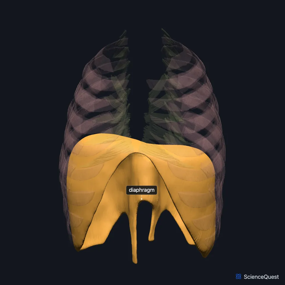
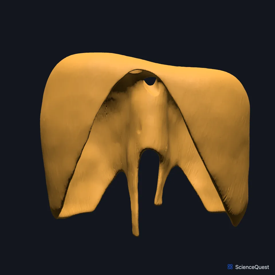
Open it in the Chest Wall Muscles Explorer See it alone in 3D
Where is the diaphragm?
Its convex upper side bulges up as the floor of the thoracic cavity, and its concave underside arches over the abdominal cavity as its roof. (OpenStax A&P 2e, 11.4; StatPearls, Thorax: Diaphragm; Gray 1918, 6c. Muscles of the Thorax)
The two halves are not level. The left sits a little lower than the right, chiefly because the liver lies under the right half, and pressure from the heart may lower the left further. (StatPearls, Thorax: Diaphragm)
What does the diaphragm attach to?
Its muscle fibers rise from the rim of the lower opening of the rib cage and converge on a central tendon at the crest of the dome. (StatPearls, Thorax: Diaphragm)
The fibers arise in three parts. The sternal part is two slips from the back of the xiphoid process of the sternum, the costal part comes from the inner surfaces of the lower six costal cartilages on each side, and the lumbar part comes from the upper lumbar vertebrae. (StatPearls, Thorax: Diaphragm)
The lumbar part has two crura. The right crus arises from the bodies of the upper three lumbar vertebrae and the discs between them, the left from the upper two, and their inner edges arch across the front of the aorta as the median arcuate ligament. (StatPearls, Thorax: Diaphragm)
What does the diaphragm do?
When it contracts it pulls its central tendon down and flattens. That deepens the thorax and lowers the pressure inside it, which draws air in. (StatPearls, Thorax: Diaphragm; OpenStax A&P 2e, 22.3)
In quiet breathing it works with the external intercostal muscles. Breathing out at rest takes no muscular effort: the lungs recoil as the diaphragm and intercostals relax. (OpenStax A&P 2e, 22.3; StatPearls, Physiology, Respiratory Drive)
When you take a deep breath and hold it, the diaphragm helps the abdominal muscles raise the pressure inside the abdomen, a move called the Valsalva maneuver. The same straining helps in urination, defecation and childbirth. (OpenStax A&P 2e, 11.4; StatPearls, Thorax: Diaphragm; Gray 1918, 6c. Muscles of the Thorax)
As it descends, pressure falls in the chest and rises in the abdomen. StatPearls calls this a thoracoabdominal pump, one that pushes blood in the inferior vena cava up into the right atrium. (StatPearls, Thorax: Diaphragm)
What passes through the diaphragm?
Three large openings carry structures between thorax and abdomen. The inferior vena cava passes through the caval opening in the central tendon at the level of T8, and the esophagus with the anterior and posterior vagal trunks passes through the esophageal hiatus at T10. (OpenStax A&P 2e, 11.4; StatPearls, Thorax: Diaphragm; FMA, Anterior vagal trunk and Posterior vagal trunk)
The aorta, thoracic duct and azygos vein pass through the aortic hiatus, which lies between the crura in front of the body of T12. (OpenStax A&P 2e, 11.4; StatPearls, Thorax: Diaphragm)
Fibers from the right crus loop around the esophagus in a sling. StatPearls says they act as a sphincter that likely helps keep stomach contents from rising into the thoracic esophagus. (StatPearls, Thorax: Diaphragm)
What lies against the diaphragm?
The fibrous pericardium around the heart is partly blended with the top of its central tendon, and the parietal pleura fuses onto the tendon too. (OpenStax A&P 2e, 11.4; StatPearls, Thorax: Diaphragm; Gray 1918, 6c. Muscles of the Thorax)
At the back, two tendinous arches complete its origin: the medial arcuate ligament (medial lumbocostal arch) over the psoas major, and the lateral arcuate ligament (lateral lumbocostal arch) over the upper quadratus lumborum, which runs out to the 12th rib. (StatPearls, Thorax: Diaphragm; FMA, Medial arcuate ligament and Lateral arcuate ligament)
Its costal part interlocks with the transversus abdominis, the deepest layer of the abdominal wall at the flanks. (StatPearls, Thorax: Diaphragm; OpenStax A&P 2e, 11.4)
Which nerves and arteries supply the diaphragm?
The right and left phrenic nerves, from the anterior rami of C3 to C5, give its motor supply. They also carry sensation from the pleura and peritoneum over its center; the lower six intercostal nerves supply sensation to its edge. (StatPearls, Thorax, Muscles and Thorax: Diaphragm)
Arteries that supply it include the musculophrenic branch of the internal thoracic artery, the superior phrenic branches of the aorta, the lower five intercostal arteries, the subcostal artery and the inferior phrenic arteries. (StatPearls, Thorax, Muscles)
How does the diaphragm form, and what if it forms with a gap?
It develops from four sources: the septum transversum, the pleuroperitoneal membranes, the mesentery of the esophagus and mesoderm of the body wall. (StatPearls, Thorax: Diaphragm)
In a congenital diaphragmatic hernia the diaphragm does not develop normally before birth, and abdominal organs pass into the chest. The Bochdalek hernia is the most common type. (StatPearls, Thorax: Diaphragm)
| Feature | Detail and source |
|---|---|
| Origin | Back of the xiphoid process; inner surfaces of the lower six costal cartilages; upper lumbar vertebrae through the two crura (StatPearls, Thorax: Diaphragm) |
| Insertion | Its own central tendon (StatPearls, Thorax: Diaphragm) |
| Action | Pulls the central tendon down in inspiration, deepening the thorax (StatPearls, Thorax: Diaphragm) |
| Nerve | Right and left phrenic nerves (C3 to C5) (StatPearls, Thorax: Diaphragm) |
| Main openings | Caval opening (T8), esophageal hiatus (T10), aortic hiatus (T12) (StatPearls, Thorax: Diaphragm) |
Touches in this model
Measured on the BodyParts3D meshes: surfaces within 2 mm of the diaphragm, nearest first, 12 of 84.
- External intercostal muscle in contact
- Innermost intercostal muscle in contact
- Internal intercostal muscle in contact
- Left transversus thoracis in contact
- Right transversus thoracis in contact
- Bronchus in contact
- Descending aorta in contact
- Eleventh thoracic vertebra in contact
- Esophagus in contact
- First lumbar vertebra in contact
- Inferior vena cava in contact
- Intervertebral disk of eleventh thoracic vertebra in contact
Sources
- OpenStax Anatomy and Physiology 2e, section 11.4, Axial Muscles of the Abdominal Wall, and Thorax. CC BY-NC-SA 4.0. Read 2026-10-25.
- StatPearls, Anatomy, Thorax: Diaphragm. Kanwal Naveen S. Bains; Sarang Kashyap; Sarah L. Lappin. Last update July 24, 2023. CC BY-NC-ND 4.0. Read 2026-10-25.
- Gray’s Anatomy of the Human Body, 20th US edition (1918), IV. Myology, 6c. The Muscles of the Thorax (1918 terms: Intercostales externi and interni for the external and internal intercostals, anterior intercostal membranes for external intercostal membranes). Henry Gray. Public domain. Read 2026-10-25.
- OpenStax Anatomy and Physiology 2e, section 22.3, The Process of Breathing. CC BY-NC-SA 4.0. Read 2026-10-25.
- StatPearls, Physiology, Respiratory Drive. Joshua E. Brinkman; Fadi Toro; Sandeep Sharma. Last update June 5, 2023. CC BY-NC-ND 4.0. Read 2026-10-25.
- Foundational Model of Anatomy, FMA6226, Anterior vagal trunk (read on EBI OLS). FMA release terms, University of Washington. Read 2026-10-25.
- Foundational Model of Anatomy, FMA6227, Posterior vagal trunk (read on EBI OLS). FMA release terms, University of Washington. Read 2026-10-25.
- Foundational Model of Anatomy, FMA58282, Medial arcuate ligament (read on EBI OLS). FMA release terms, University of Washington. Read 2026-10-25.
- Foundational Model of Anatomy, FMA58283, Lateral arcuate ligament (read on EBI OLS). FMA release terms, University of Washington. Read 2026-10-25.
- StatPearls, Anatomy, Thorax, Muscles. Alex Tang; Bruno Bordoni. Last update July 24, 2023. CC BY-NC-ND 4.0. Read 2026-10-25.
The values are facts and free to use; this page’s selection and presentation are © 2026 ScienceQuest.