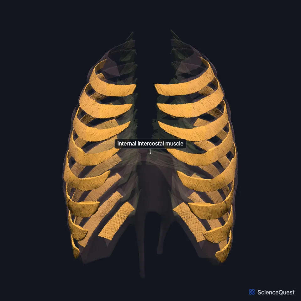
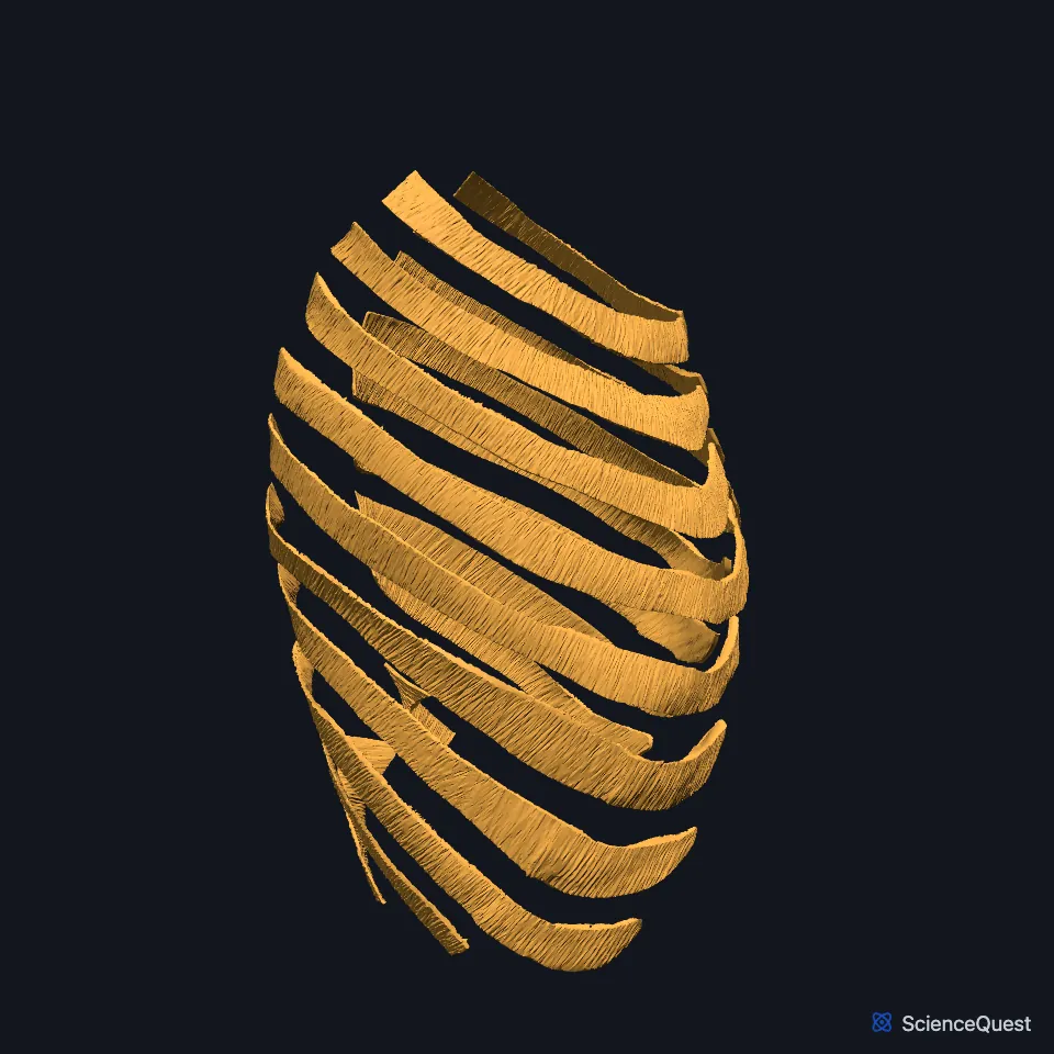
Internal intercostal muscle
The internal intercostal muscles form the middle of the three muscle layers between the ribs. The 11 pairs lie just under the external intercostals and draw the ribs together when you breathe out hard. (OpenStax A&P 2e, 11.4; StatPearls, Thorax, Muscles)
Open it in the Chest Wall Muscles Explorer See it alone in 3D
Where are the internal intercostal muscles?
They run from the sternum at the front to the angles of the ribs at the back. Behind that, a thin internal intercostal membrane takes their place. (StatPearls, Thorax, Muscles)
Near the sternum they lie with no external intercostal fibers over them, because the external intercostals stop at the costal cartilages and continue forward as a thin membrane. The innermost intercostals form the deepest layer. (OpenStax A&P 2e, 11.4; StatPearls, Thorax, Muscles; Gray 1918, 6c. Muscles of the Thorax)
What do the internal intercostals attach to?
In StatPearls each arises from the outer part of the costal groove of the rib above and inserts on the upper border of the rib below. Its fibers cross those of the external intercostals at right angles. (StatPearls, Thorax, Muscles)
What do the internal intercostals do?
Their fiber direction lets them pull the ribs down and shrink the thorax in forced expiration. (StatPearls, Thorax, Muscles)
Breathing out at rest is passive. In hard breathing, as in exercise or singing, the internal intercostals work as accessory muscles, and the abdominal muscles push the abdominal organs up toward the diaphragm. (OpenStax A&P 2e, 22.3; StatPearls, Physiology, Respiratory Drive, Thorax, Muscles and Anterolateral Abdominal Wall Muscles)
The innermost intercostals work with them as synergists. (OpenStax A&P 2e, 11.4; StatPearls, Thorax, Muscles)
Which nerves and vessels run with the internal intercostals?
Each intercostal nerve runs in the groove under its rib, between the internal intercostal and the innermost intercostal, and the intercostal arteries and veins travel in the same plane. (StatPearls, Intercostal Nerves and Intercostal Neurovascular Bundle)
Along most of the groove the vein lies highest, the artery below it and the nerve lowest. A collateral branch of the nerve runs along the upper border of the rib below. (StatPearls, Intercostal Nerves and Intercostal Neurovascular Bundle)
Muscular branches of the intercostal nerves supply the intercostal muscles. The internal thoracic artery runs down between the internal intercostals and transversus thoracis, behind the upper six costal cartilages. (StatPearls, Intercostal Nerves and Abdominal Wall)
Where the sources differ
OpenStax’s table of thorax muscles gives the internal intercostal’s origin as the rib below and its insertion as the rib above. StatPearls describes the reverse: from the costal groove of the rib above to the upper border of the rib below. (OpenStax A&P 2e, 11.4; StatPearls, Thorax, Muscles)
| Feature | Detail and source |
|---|---|
| Origin | Outer part of the costal groove of the rib above (StatPearls, Thorax, Muscles) |
| Insertion | Upper border of the rib below (StatPearls, Thorax, Muscles) |
| Action | Pull the ribs down in forced expiration (StatPearls, Thorax, Muscles) |
| Nerve | Intercostal nerves (anterior rami of T1 to T11) (StatPearls, Thorax, Muscles) |
Touches in this model
Measured on the BodyParts3D meshes: surfaces within 2 mm of the internal intercostal muscle, nearest first, 12 of 75.
- Diaphragm in contact
- External intercostal muscle in contact
- Innermost intercostal muscle in contact
- Left transversus thoracis in contact
- Right transversus thoracis in contact
- Abdominal part of left pectoralis major in contact
- Abdominal part of right pectoralis major in contact
- Body of sternum in contact
- Left costal cartilage in contact
- Left eighth rib in contact
- Left eleventh rib in contact
- Left external oblique in contact
Sources
- OpenStax Anatomy and Physiology 2e, section 11.4, Axial Muscles of the Abdominal Wall, and Thorax. CC BY-NC-SA 4.0. Read 2026-10-25.
- StatPearls, Anatomy, Thorax, Muscles. Alex Tang; Bruno Bordoni. Last update July 24, 2023. CC BY-NC-ND 4.0. Read 2026-10-25.
- Gray’s Anatomy of the Human Body, 20th US edition (1918), IV. Myology, 6c. The Muscles of the Thorax (1918 terms: Intercostales externi and interni for the external and internal intercostals, anterior intercostal membranes for external intercostal membranes). Henry Gray. Public domain. Read 2026-10-25.
- OpenStax Anatomy and Physiology 2e, section 22.3, The Process of Breathing. CC BY-NC-SA 4.0. Read 2026-10-25.
- StatPearls, Physiology, Respiratory Drive. Joshua E. Brinkman; Fadi Toro; Sandeep Sharma. Last update June 5, 2023. CC BY-NC-ND 4.0. Read 2026-10-25.
- StatPearls, Anatomy, Anterolateral Abdominal Wall Muscles. Matthew A. Varacallo; Stephanie Scharbach; Mohammed A. Al-Dhahir. Last update July 24, 2023. CC BY-NC-ND 4.0. Read 2026-10-25.
- StatPearls, Anatomy, Thorax, Intercostal Nerves. Niklas L. Glenesk; Sajedur Rahman; Peter P. Lopez. Last update May 22, 2023. CC BY-NC-ND 4.0. Read 2026-10-25.
- StatPearls, Anatomy, Thoracotomy and the Collateral Intercostal Neurovascular Bundle. Noel T. Boaz; Raymond L. Bernor; Keiko Meshida; Forshing Lui. Last update September 26, 2022. CC BY-NC-ND 4.0. Read 2026-10-25.
- StatPearls, Anatomy, Abdomen and Pelvis: Abdominal Wall. William Flynn; Paula Vickerton. Last update July 24, 2023. CC BY-NC-ND 4.0. Read 2026-10-25.
The values are facts and free to use; this page’s selection and presentation are © 2026 ScienceQuest.