External oblique
The external oblique is the outermost and largest of the three flat muscles of the abdominal wall. Its fibers run down and toward the midline, the way your fingers point when you slide them into your front pockets. (StatPearls, Anterolateral Abdominal Wall Muscles; OpenStax A&P 2e, 11.4)
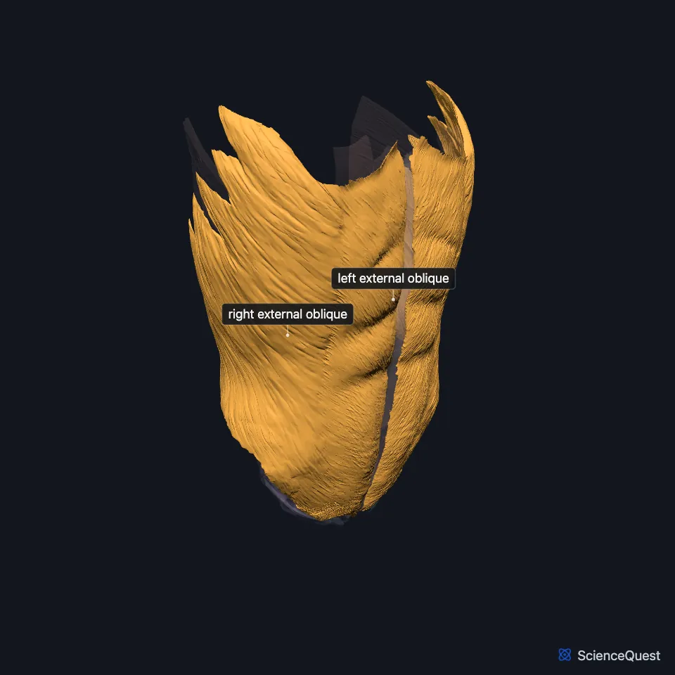
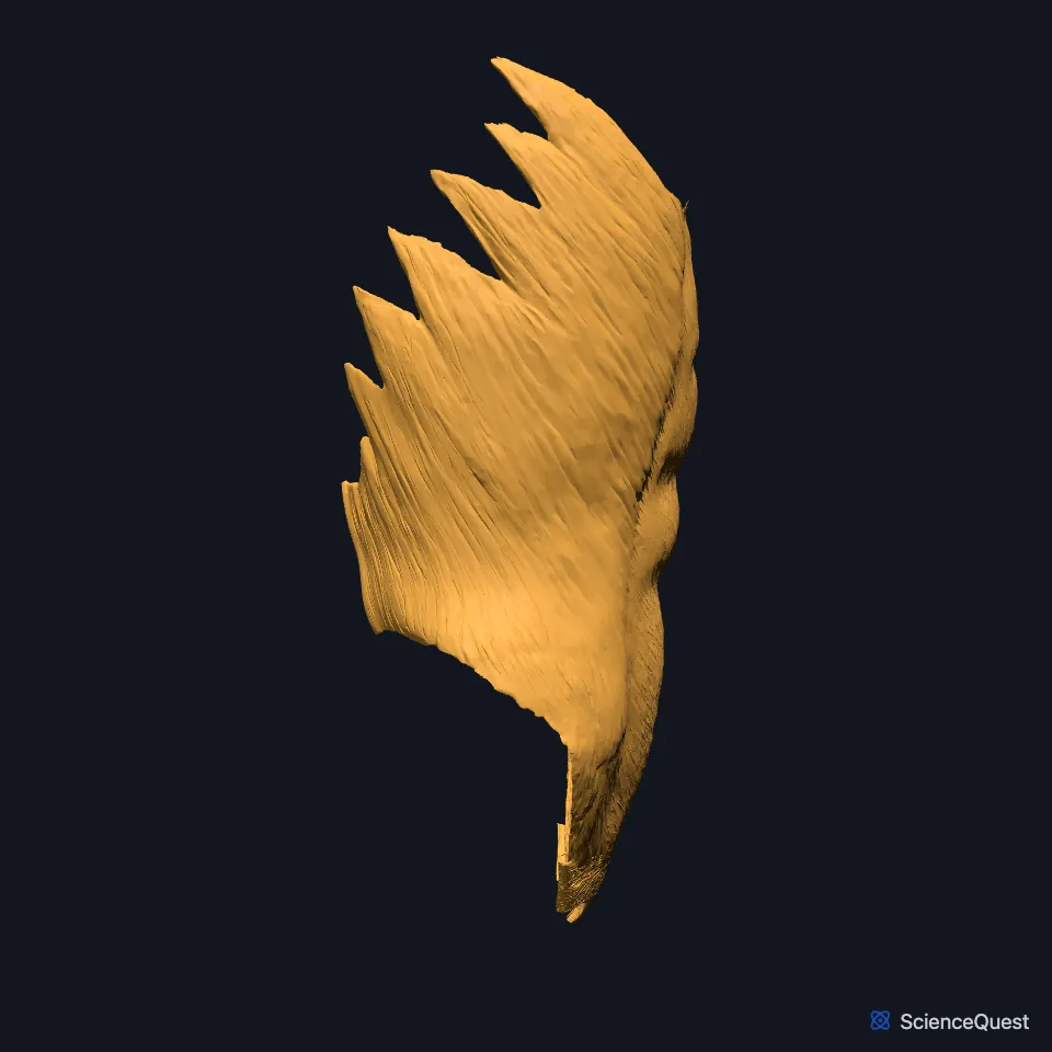
Open it in the Abdominal Muscles Explorer See it alone in 3D
Where is the external oblique?
It is the outermost, largest and thickest of the three flat muscles of the anterolateral abdominal wall. At about the midclavicular line its fleshy part ends in an aponeurosis, a flat sheet of tendon, which passes in front of the rectus abdominis to the linea alba. (StatPearls, Abdominal Wall and Anterolateral Abdominal Wall)
Its lowest edge folds back and up to form the inguinal ligament, which runs from the anterior superior iliac spine to the pubic tubercle. (StatPearls, Anterolateral Abdominal Wall and Abdominal Wall)
What does the external oblique attach to?
It arises from the lower ribs and inserts on the front half of the iliac crest. StatPearls also calls it the thickest of the three flat muscles. (StatPearls, Anterolateral Abdominal Wall)
What does the external oblique do?
With the internal oblique, it rotates the vertebral column and bends it to the side: twisting at the waist. (StatPearls, Abdominal Wall; OpenStax A&P 2e, 11.4)
Its fibers run down and toward the midline, the internal oblique’s run up and toward the midline, and the transversus abdominis runs across. Together these flat muscles flex and rotate the trunk and help shield the abdominal organs from injury. (OpenStax A&P 2e, 11.4; StatPearls, Anterolateral Abdominal Wall Muscles)
Because the fibers of the three flat muscles criss-cross and interlink, they strengthen the wall and reduce the risk of herniation. In forced expiration the obliques contract and push the abdominal organs up against the diaphragm. (StatPearls, Anterolateral Abdominal Wall Muscles; OpenStax A&P 2e, 22.3)
What lies under the external oblique, and what supplies it?
It forms the outer layer of the flank, with the internal oblique beneath it and the transversus abdominis deepest. (OpenStax A&P 2e, 11.4; StatPearls, Anterolateral Abdominal Wall)
The anterior rami of T7 to T12, the lower intercostal nerves (T7 to T11) and the subcostal nerve (T12), supply the anterolateral wall, running forward between the internal oblique and the transversus abdominis. (StatPearls, Anterolateral Abdominal Wall, Thorax, Muscles, Abdominal Wall and Intercostal Nerves)
Part of the pectoralis major arises from its aponeurosis. (StatPearls, Thorax, Muscles)
Where the sources differ
StatPearls’ abdominal wall article has it arising from the fifth to twelfth ribs. Its anterolateral abdominal wall article says the lower seven ribs. (StatPearls, Abdominal Wall and Anterolateral Abdominal Wall)
OpenStax’s table, which groups the two obliques, gives their origin as ribs 5 to 12 and the ilium and their insertion as ribs 7 to 10, the linea alba and the ilium. StatPearls’ anterolateral abdominal wall article inserts the external oblique on the front half of the iliac crest and, through its aponeurosis, the linea alba. (OpenStax A&P 2e, 11.4; StatPearls, Anterolateral Abdominal Wall)
| Feature | Detail and source |
|---|---|
| Origin | Lower ribs: the fifth to twelfth, or the lower seven (StatPearls, Abdominal Wall and Anterolateral Abdominal Wall) |
| Insertion | Front half of the iliac crest; linea alba through its aponeurosis (StatPearls, Anterolateral Abdominal Wall) |
| Action | Rotates the trunk and bends it to the side, with the internal oblique (StatPearls, Abdominal Wall) |
| Nerve | Anterior rami of T7 to T12 (intercostal nerves T7 to T11, subcostal nerve T12) (StatPearls, Anterolateral Abdominal Wall and Thorax, Muscles) |
Touches in this model
Measured on the BodyParts3D meshes: surfaces within 2 mm of the external oblique, nearest first, 12 of 60.
- Left inguinal ligament in contact
- Left internal oblique in contact
- Left rectus abdominis in contact
- Linea alba in contact
- Right inguinal ligament in contact
- Right internal oblique in contact
- Right rectus abdominis in contact
- Abdominal part of left pectoralis major in contact
- Abdominal part of right pectoralis major in contact
- Body of sternum in contact
- External intercostal muscle in contact
- Internal intercostal muscle in contact
Sources
- StatPearls, Anatomy, Anterolateral Abdominal Wall Muscles. Matthew A. Varacallo; Stephanie Scharbach; Mohammed A. Al-Dhahir. Last update July 24, 2023. CC BY-NC-ND 4.0. Read 2026-10-25.
- OpenStax Anatomy and Physiology 2e, section 11.4, Axial Muscles of the Abdominal Wall, and Thorax. CC BY-NC-SA 4.0. Read 2026-10-25.
- StatPearls, Anatomy, Abdomen and Pelvis: Abdominal Wall. William Flynn; Paula Vickerton. Last update July 24, 2023. CC BY-NC-ND 4.0. Read 2026-10-25.
- StatPearls, Anatomy, Abdomen and Pelvis: Anterolateral Abdominal Wall. Kevin Seeras; Ryan N. Qasawa; Ricky Ju; Shivana Prakash. Last update July 24, 2023. CC BY-NC-ND 4.0. Read 2026-10-25.
- OpenStax Anatomy and Physiology 2e, section 22.3, The Process of Breathing. CC BY-NC-SA 4.0. Read 2026-10-25.
- StatPearls, Anatomy, Thorax, Muscles. Alex Tang; Bruno Bordoni. Last update July 24, 2023. CC BY-NC-ND 4.0. Read 2026-10-25.
- StatPearls, Anatomy, Thorax, Intercostal Nerves. Niklas L. Glenesk; Sajedur Rahman; Peter P. Lopez. Last update May 22, 2023. CC BY-NC-ND 4.0. Read 2026-10-25.
The values are facts and free to use; this page’s selection and presentation are © 2026 ScienceQuest.