Rectus abdominis
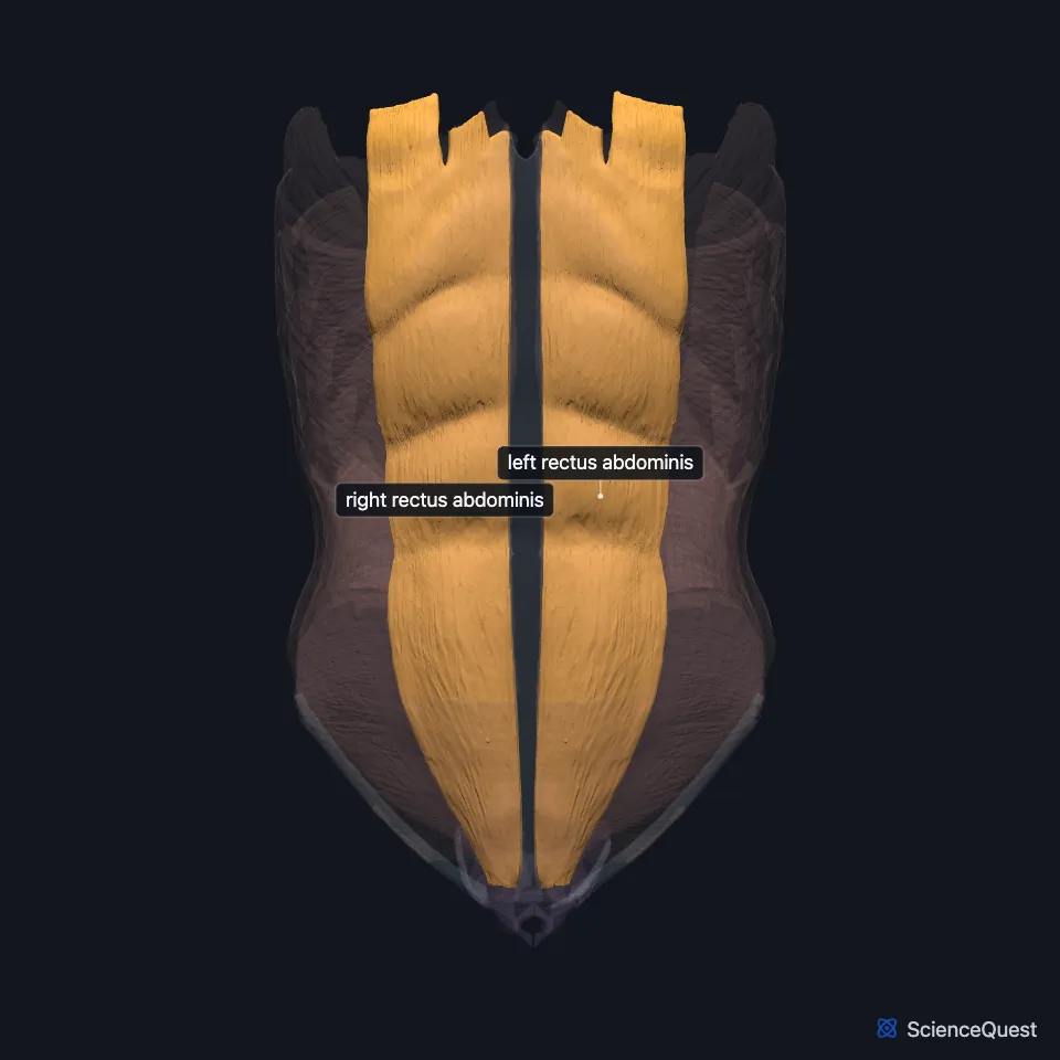
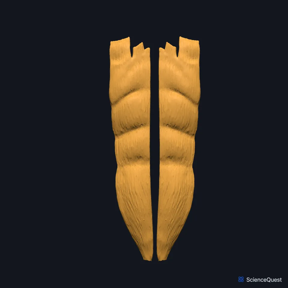
The rectus abdominis is a long, narrow muscle that runs up the front of the abdomen beside the midline, from the pubis to the fifth to seventh costal cartilages. It is a powerful flexor of the vertebral column, commonly called the sit-up muscle. (StatPearls, Abdominal Wall; OpenStax A&P 2e, 11.4)
Open it in the Abdominal Muscles Explorer See it alone in 3D
Where is the rectus abdominis?
The right and left muscles lie either side of the linea alba, the fibrous seam in the midline where the aponeuroses of the flat abdominal muscles of the two sides meet. It runs from the xiphoid process to the pubic symphysis. (OpenStax A&P 2e, 11.4; StatPearls, Abdominal Wall; Gray 1918, 6d. Muscles of the Abdomen)
Its outer border is the linea semilunaris. Low on its front, inside the same sheath, sits the small triangular pyramidalis, which is absent in 20% of people. (StatPearls, Anterolateral Abdominal Wall Muscles, Abdominal Wall and Rectus Sheath)
What gives the rectus abdominis its six-pack?
Three tendinous intersections, bands of collagen, cross each muscle and divide it into four segments. They are tethered to the front wall of the rectus sheath, and in athletes they can show as a six-pack. (StatPearls, Abdominal Wall; OpenStax A&P 2e, 11.4)
What is the rectus sheath?
The muscle runs inside the rectus sheath, which the aponeuroses of the external oblique, internal oblique and transversus abdominis build. The sheath reaches from the costal margin and the cartilages of ribs 5 to 7 down to the pubic crest. (StatPearls, Rectus Sheath)
Above the arcuate line, the external oblique’s layer and half of the internal oblique’s pass in front of the muscle, while the other half, the transversus abdominis layer and the transversalis fascia pass behind it. Below the line, all three layers pass in front, and only the transversalis fascia lies behind the muscle. (StatPearls, Rectus Sheath)
StatPearls’ rectus sheath article says the change in the fibers at the arcuate line leaves the wall weaker where that line meets the outer edge of the sheath, and calls a hernia there a Spigelian hernia. (StatPearls, Rectus Sheath)
What does the rectus abdominis do?
It flexes the lumbar spine. It also compresses the abdominal organs and steadies the pelvis during walking. (StatPearls, Rectus Sheath and Anterolateral Abdominal Wall Muscles)
Contracting with the other abdominal muscles, it raises the pressure in the abdomen and chest needed to sneeze, cough, vomit and defecate, and may help steady the trunk when you lift heavy loads. (StatPearls, Abdominal Wall)
Which nerves and arteries supply the rectus abdominis?
The thoracoabdominal nerves from T7 to T11 and the subcostal nerve (T12) supply it. (StatPearls, Rectus Sheath)
The superior epigastric artery, from the internal thoracic artery, and the inferior epigastric artery, from the external iliac artery, supply it, and the two join each other to varying degrees. Small branches of the lower six intercostal arteries also contribute. (StatPearls, Rectus Sheath and Anterolateral Abdominal Wall)
Inside the sheath the superior epigastric artery runs behind the muscle, in front of the sheath’s back wall. The inferior epigastric artery climbs on the deep surface of the muscle and enters the sheath at the arcuate line, then runs on behind the muscle. (StatPearls, Anterolateral Abdominal Wall and Abdominal Wall)
Where the sources differ
StatPearls articles put the arcuate line, where the back wall of the sheath ends, at different heights: about midway between the umbilicus and the pubic symphysis, a third of the distance between the umbilicus and the pubis, or just below the level of the umbilicus. (StatPearls, Anterolateral Abdominal Wall Muscles, Rectus Sheath and Anterolateral Abdominal Wall)
OpenStax’s table gives its insertion as the sternum and ribs 5 and 7. StatPearls gives the fifth to seventh costal cartilages. (OpenStax A&P 2e, 11.4; StatPearls, Abdominal Wall)
StatPearls’ rectus sheath article puts the weak point behind a Spigelian hernia where the arcuate line meets the outer edge of the sheath. Its abdominal wall article puts Spigelian hernias on the semilunar line, the outer border of the rectus abdominis and its sheath, usually above the arcuate line. (StatPearls, Rectus Sheath and Abdominal Wall)
| Feature | Detail and source |
|---|---|
| Origin | Pubic symphysis and pubic crest (StatPearls, Abdominal Wall; OpenStax A&P 2e, 11.4) |
| Insertion | Fifth to seventh costal cartilages (StatPearls, Abdominal Wall) |
| Action | Flexes the vertebral column; compresses the abdominal organs (StatPearls, Abdominal Wall and Anterolateral Abdominal Wall Muscles) |
| Nerve | Thoracoabdominal nerves (T7 to T11) and subcostal nerve (T12) (StatPearls, Rectus Sheath) |
Touches in this model
Measured on the BodyParts3D meshes: surfaces within 2 mm of the rectus abdominis, nearest first, 12 of 37.
- Left external oblique in contact
- Left internal oblique in contact
- Left pyramidalis in contact
- Left transversus abdominis in contact
- Linea alba in contact
- Right external oblique in contact
- Right internal oblique in contact
- Right pyramidalis in contact
- Right transversus abdominis in contact
- Abdominal part of left pectoralis major in contact
- Abdominal part of right pectoralis major in contact
- Internal intercostal muscle in contact
Sources
- StatPearls, Anatomy, Abdomen and Pelvis: Abdominal Wall. William Flynn; Paula Vickerton. Last update July 24, 2023. CC BY-NC-ND 4.0. Read 2026-10-25.
- OpenStax Anatomy and Physiology 2e, section 11.4, Axial Muscles of the Abdominal Wall, and Thorax. CC BY-NC-SA 4.0. Read 2026-10-25.
- Gray’s Anatomy of the Human Body, 20th US edition (1918), IV. Myology, 6d. The Muscles and Fasciae of the Abdomen. Henry Gray. Public domain. Read 2026-10-25.
- StatPearls, Anatomy, Anterolateral Abdominal Wall Muscles. Matthew A. Varacallo; Stephanie Scharbach; Mohammed A. Al-Dhahir. Last update July 24, 2023. CC BY-NC-ND 4.0. Read 2026-10-25.
- StatPearls, Anatomy, Abdomen and Pelvis, Rectus Sheath. Karlin E. Sevensma; Logan Leavitt; Kerent D. Pihl. Last update July 24, 2023. CC BY-NC-ND 4.0. Read 2026-10-25.
- StatPearls, Anatomy, Abdomen and Pelvis: Anterolateral Abdominal Wall. Kevin Seeras; Ryan N. Qasawa; Ricky Ju; Shivana Prakash. Last update July 24, 2023. CC BY-NC-ND 4.0. Read 2026-10-25.
The values are facts and free to use; this page’s selection and presentation are © 2026 ScienceQuest.