Upper Limb Muscles Explorer
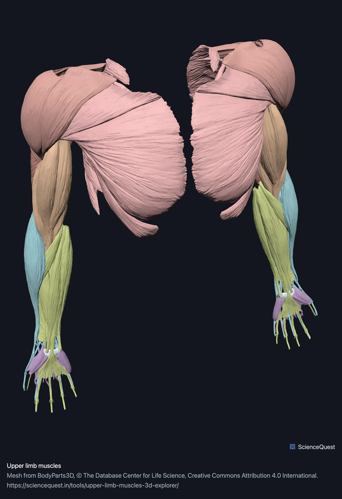
Turn 114 muscle meshes of both upper limbs in 3D: pectoral girdle, rotator cuff, arm and forearm compartments, and the intrinsic muscles of the hand.
Visualiser
Upper limb muscles, 116 structures
Checking this device for a stored copy.
Start here
- Hide the Pectoral girdle band It is the outermost layer on the front of the chest. Hidden, subscapularis, which lines the front of the shoulder blade, comes into view. The other three cuff muscles lie on the back of the blade, and the deltoid capping the shoulder stays visible because it is in the cuff band.
- Click Posterior forearm, then Back Eleven muscles a side in this band, stacked in two layers, and from the front you can see almost none of them. Anconeus, which many texts count as a twelfth muscle of the compartment, is in the Posterior arm band.
- Select the long head of biceps brachii Its tendon runs inside the shoulder joint capsule, over the head of the humerus, while the short head passes in front of the joint from the coracoid process. That intra-articular course is why the long head matters for shoulder pain. Two separate meshes let you follow each head up to the shoulder rather than take it on trust.
Muscle, and the flexor retinaculum at each wrist. No bone, nerves or vessels, and no tendon sheaths. Most tendons are part of their muscle belly rather than separate, so the long flexor tendons cannot be followed into the fingers individually.
- 116 structures
- 3313k triangles
- 17000 KB, on request
- 0.00526 mm worst error per axis
- 12 pectoral girdle
- 16 rotator cuff, deltoid and teres major
- 8 anterior arm
- 8 posterior arm
- 22 anterior forearm
- 24 posterior forearm
- 24 hand
- 2 retinaculum
Where this comes from
Segmented from imaging of one adult male body, not drawn by an artist, so the shapes are measurements. Two consequences worth knowing: it is one person’s anatomy rather than an average, and faint stepping on some surfaces is the resolution of the original scan rather than a rendering fault.
Meshes from BodyParts3D, © The Database Center for Life Science, under Creative Commons Attribution 4.0 International. Names and identifiers are the dataset’s own: Foundational Model of Anatomy identifiers and English labels, as its parts list gives them. Converted meshes are shared under the same licence.
Changes made: Meshes were converted from binary STL to an indexed mesh with 16-bit quantised vertex positions, and surface normals were recomputed from the geometry. No vertex was moved other than by that quantisation, whose worst-case error on any one coordinate is reported on the page.
Latin terms for 60 of the 116 structures come from English Wikipedia anatomy infoboxes, under Creative Commons Attribution-ShareAlike 4.0, joined by Wikidata property P1402, Foundational Model of Anatomy identifier. Only a term a source states for that structure is used, never one constructed from a family name, which is why the rest carry no Latin rather than a guess.
All 116 structures
Every structure the model contains, group by group and alphabetically within each. Each name opens the viewer with that structure selected, so a link can point at one structure rather than at the whole region.
Pectoral girdle 12
- abdominal part of left pectoralis major muscle
- abdominal part of right pectoralis major muscle
- clavicular part of left pectoralis major muscle
- clavicular part of right pectoralis major muscle
- left pectoralis minor musculus pectoralis minor muscle
- left serratus anterior musculus serratus anterior, serratus lateralis muscle
- left subclavius musculus subclavius muscle
- right pectoralis minor musculus pectoralis minor muscle
- right serratus anterior musculus serratus anterior, serratus lateralis muscle
- right subclavius musculus subclavius muscle
- sternocostal part of left pectoralis major muscle
- sternocostal part of right pectoralis major muscle
Rotator cuff, deltoid and teres major 16
- acromial part of left deltoid muscle
- acromial part of right deltoid muscle
- clavicular part of left deltoid muscle
- clavicular part of right deltoid muscle
- left infraspinatus muscle musculus infraspinatus muscle
- left subscapularis musculus subscapularis muscle
- left supraspinatus musculus supraspinatus muscle
- left teres major musculus teres major muscle
- left teres minor musculus teres minor muscle
- right infraspinatus muscle musculus infraspinatus muscle
- right subscapularis musculus subscapularis muscle
- right supraspinatus musculus supraspinatus muscle
- right teres major musculus teres major muscle
- right teres minor musculus teres minor muscle
- spinal part of left deltoid muscle
- spinal part of right deltoid muscle
Anterior arm 8
- left brachialis musculus brachialis muscle
- left coracobrachialis musculus coracobrachialis muscle
- long head of left biceps brachii muscle
- long head of right biceps brachii muscle
- right brachialis musculus brachialis muscle
- right coracobrachialis musculus coracobrachialis muscle
- short head of left biceps brachii muscle
- short head of right biceps brachii muscle
Posterior arm 8
- lateral head of left triceps brachii muscle
- lateral head of right triceps brachii muscle
- left anconeus musculus anconeus, musculus anconaeus muscle
- long head of left triceps brachii muscle
- long head of right triceps brachii muscle
- medial head of left triceps brachii muscle
- medial head of right triceps brachii muscle
- right anconeus musculus anconeus, musculus anconaeus muscle
Anterior forearm 22
- humeral head of left flexor carpi ulnaris muscle
- humeral head of left pronator teres muscle
- humeral head of right flexor carpi ulnaris muscle
- humeral head of right pronator teres muscle
- humeroulnar head of left flexor digitorum superficialis muscle
- humeroulnar head of right flexor digitorum superficialis muscle
- left flexor carpi radialis musculus flexor carpi radialis muscle
- left flexor digitorum profundus musculus flexor digitorum profundus muscle
- left flexor pollicis longus musculus flexor pollicis longus muscle
- left palmaris longus musculus palmaris longus muscle
- left pronator quadratus musculus pronator quadratus muscle
- radial head of left flexor digitorum superficialis muscle
- radial head of right flexor digitorum superficialis muscle
- right flexor carpi radialis musculus flexor carpi radialis muscle
- right flexor digitorum profundus musculus flexor digitorum profundus muscle
- right flexor pollicis longus musculus flexor pollicis longus muscle
- right palmaris longus musculus palmaris longus muscle
- right pronator quadratus musculus pronator quadratus muscle
- ulnar head of left flexor carpi ulnaris muscle
- ulnar head of left pronator teres muscle
- ulnar head of right flexor carpi ulnaris muscle
- ulnar head of right pronator teres muscle
Posterior forearm 24
- humeral head of left extensor carpi ulnaris muscle
- humeral head of right extensor carpi ulnaris muscle
- left abductor pollicis longus musculus abductor pollicis longus muscle
- left brachioradialis musculus brachioradialis muscle
- left extensor carpi radialis brevis musculus extensor carpi radialis brevis muscle
- left extensor carpi radialis longus musculus extensor carpi radialis longus muscle
- left extensor digiti minimi musculus extensor digiti minimi muscle
- left extensor digitorum musculus extensor digitorum muscle
- left extensor indicis musculus extensor indicis muscle
- left extensor pollicis brevis musculus extensor pollicis brevis muscle
- left extensor pollicis longus musculus extensor pollicis longus muscle
- left supinator musculus supinator muscle
- right abductor pollicis longus musculus abductor pollicis longus muscle
- right brachioradialis musculus brachioradialis muscle
- right extensor carpi radialis brevis musculus extensor carpi radialis brevis muscle
- right extensor carpi radialis longus musculus extensor carpi radialis longus muscle
- right extensor digiti minimi musculus extensor digiti minimi muscle
- right extensor digitorum musculus extensor digitorum muscle
- right extensor indicis musculus extensor indicis muscle
- right extensor pollicis brevis musculus extensor pollicis brevis muscle
- right extensor pollicis longus musculus extensor pollicis longus muscle
- right supinator musculus supinator muscle
- ulnar head of left extensor carpi ulnaris muscle
- ulnar head of right extensor carpi ulnaris muscle
Hand 24
- abductor digiti minimi of left hand musculus abductor digiti minimi manus muscle
- abductor digiti minimi of right hand musculus abductor digiti minimi manus muscle
- deep head of left flexor pollicis brevis muscle
- deep head of right flexor pollicis brevis muscle
- flexor digiti minimi brevis of left hand musculus flexor digiti minimi brevis manus muscle
- flexor digiti minimi brevis of right hand musculus flexor digiti minimi brevis manus muscle
- left abductor pollicis brevis musculus abductor pollicis brevis muscle
- left opponens pollicis musculus opponens pollicis muscle
- oblique head of left adductor pollicis muscle
- oblique head of right adductor pollicis muscle
- opponens digiti minimi of left hand muscle
- opponens digiti minimi of right hand muscle
- right abductor pollicis brevis musculus abductor pollicis brevis muscle
- right opponens pollicis musculus opponens pollicis muscle
- set of dorsal interossei of left hand muscle
- set of dorsal interossei of right hand muscle
- set of lumbricals of left hand muscle
- set of lumbricals of right hand muscle
- set of palmar interossei of left hand muscle
- set of palmar interossei of right hand muscle
- superficial head of left flexor pollicis brevis muscle
- superficial head of right flexor pollicis brevis muscle
- transverse head of left adductor pollicis muscle
- transverse head of right adductor pollicis muscle
Retinaculum 2
- flexor retinaculum of left wrist other tissue
- flexor retinaculum of right wrist other tissue
Citing this tool
Last updated . Add the date you accessed it as well, which a citation of a page that can change asks for. If a specific view matters, cite the page address as it stands in your browser instead: it records what is selected and shown.
Compartments, not individual muscles
The forearm has an anterior and a posterior compartment, each with its own nerve supply, and the arm the same: the radial nerve behind in both, the musculocutaneous in front of the arm, and the median and ulnar in front of the forearm. That is the division that predicts what happens when a nerve is injured, so it is the division the bands use. Hiding the anterior forearm to see the posterior one behind it is one click rather than eleven.
Within a compartment the model keeps the dataset’s own subdivisions, which are finer than most atlases: pronator teres and both carpi ulnaris muscles as their humeral and ulnar heads, and flexor digitorum superficialis as its humeroulnar and radial heads.
Five muscles are deliberately not here
Trapezius in three parts, latissimus dorsi, levator scapulae and both rhomboids. The dataset files them under the upper limb because they move the shoulder girdle, and under the back as well. They ship with the back model.
The pectoral group goes the other way: pectoralis major in three parts, pectoralis minor, serratus anterior and subclavius are claimed by both the upper limb and the thorax, and they are here, because what they do is move the arm. Each overlapping muscle has exactly one home, and the build asserts it.
One structure is not a muscle
The flexor retinaculum of the wrist, one per side, is the fibrous band roofing the carpal tunnel. It is tagged as other tissue, which is why 114 of the 116 structures are muscle meshes. It is here because the long flexor tendons pass beneath it, and that space is the carpal tunnel.
Common questions
Why is the trapezius not in here?
Because it ships with the back, and it can only ship once. The dataset files trapezius, latissimus dorsi, levator scapulae and both rhomboids under the upper limb, because what they do is move the shoulder girdle, and it files them under the back as well. Both are correct. They are in the back model because that is where a reader goes looking for them, and this region excludes those five muscles, seven meshes a side, by name, so neither model claims a muscle the other has.
Why are the pectorals here rather than with the chest?
The same overlap resolved the other way. Pectoralis major in three parts, pectoralis minor, serratus anterior and subclavius sit under both the upper limb and the thorax. They are here because what they do is move the arm, which leaves the chest wall model as the wall proper: diaphragm, intercostals, levatores costarum and transversus thoracis.
Why are so many muscles split into heads?
Because the dataset publishes them that way, and it is usually the more useful division. Biceps brachii arrives as its long and short heads, triceps as all three, pronator teres and both carpi ulnaris muscles as their humeral and ulnar heads, and flexor digitorum superficialis as its humeroulnar and radial heads. That is what lets you follow each head of biceps to its own origin: both cross the shoulder as well as the elbow, but the long head’s tendon runs through the joint itself, over the head of the humerus, while the short head passes in front of it from the coracoid process. That difference is the whole reason the distinction matters.
Is anything here not a muscle?
One structure per side: the flexor retinaculum of the wrist, which is the fibrous band that roofs the carpal tunnel. It is tagged as other tissue and left out of the muscle count, which is why the figures read 114 muscle meshes within 116 structures. It is in the model because the tendons of the forearm flexors pass under it, and that relationship is the carpal tunnel.