Brain Explorer
Free 3D brain anatomy: gyri by lobe, hippocampus and limbic structures, basal ganglia, thalamus, brainstem, cerebellum and ventricles, each named.
Visualiser
Brain, 101 structures
Checking this device for a stored copy.
Start here
- Hide every cortex band, then click Ventricles The fastest orientation in the whole model. The ventricular cast is the most recognisable shape a brain has, and once it is framed everything else can be placed against it.
- Hide the Cerebellum It arrives as a single mesh, 12 percent of the model’s triangles on its own, and it sits directly over the brainstem, so hiding it is both what exposes the pons and medulla and what makes the model lighter to turn.
- Select the left hippocampus, then turn on Isolate selection Pulls one structure out of 101. Worth doing for the hippocampus and amygdala in particular, since neither can be seen at all from outside the brain.
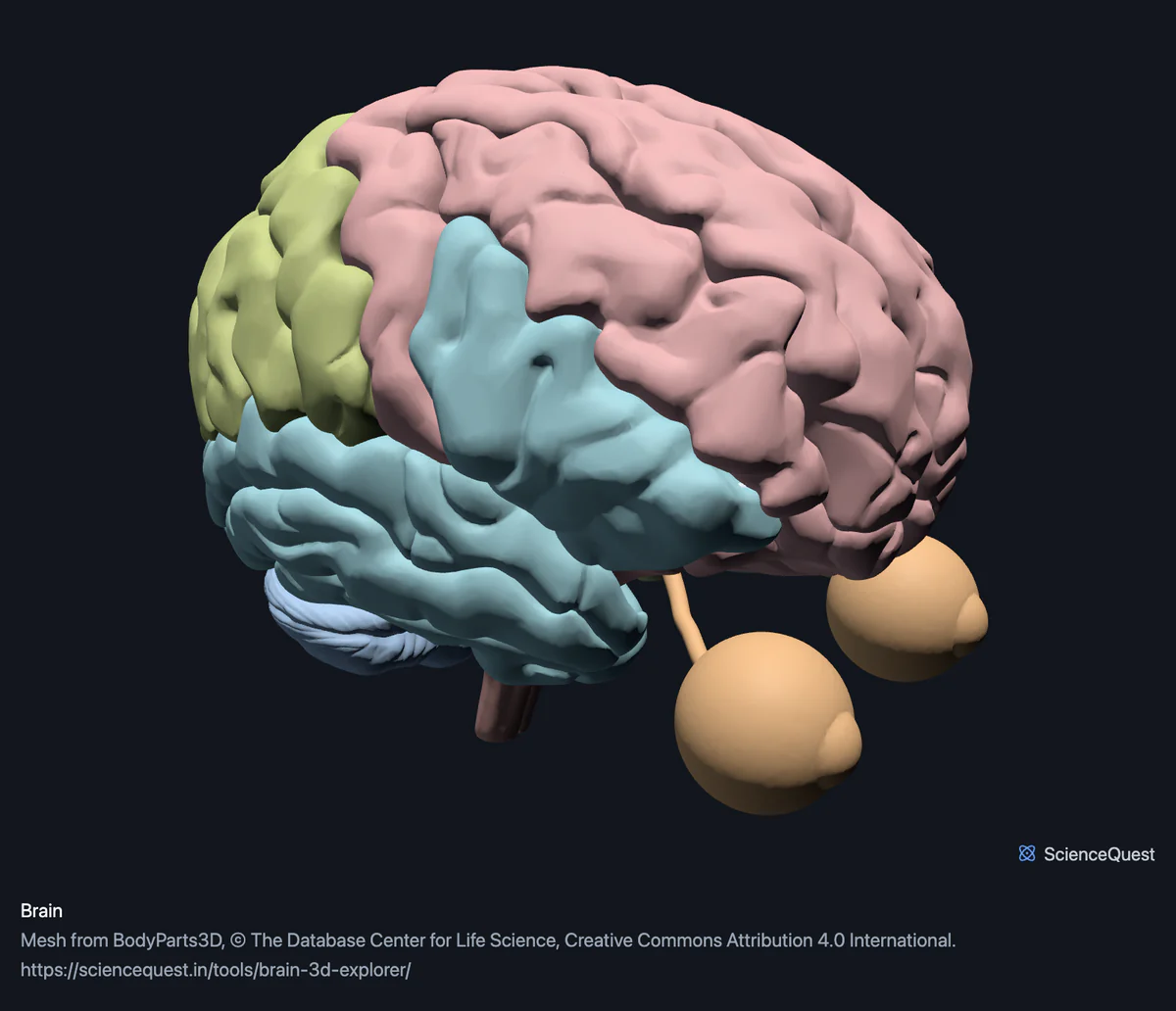
Brain, pituitary and eyeballs. No spinal cord surface, only its central canal, and no peripheral nerves apart from the optic nerves. No other cranial nerves, no blood vessels, no dura or arachnoid, and no cerebellar folia or deep nuclei: the cerebellum is one mesh. Both eyeballs arrive as one mesh named eyeball, which is why the list has a single row for two globes, and nothing else of the eye is here: no lens, retina or extraocular muscle.
- 101 structures
- 2064k triangles
- 10300 KB, on request
- 0.0013 mm worst error per axis
- 7 frontal cortex
- 8 parietal cortex
- 14 temporal cortex and insula
- 2 occipital lobe
- 20 limbic and midline structures
- 15 basal ganglia, thalamus and hypothalamus
- 13 brainstem
- 1 cerebellum
- 4 white matter
- 9 ventricles and choroid plexus
- 2 pituitary and pineal
- 6 optic pathway
Where this comes from
Segmented from imaging of one adult male body, not drawn by an artist, so the shapes are measurements. Two consequences worth knowing: it is one person’s anatomy rather than an average, and faint stepping on some surfaces is the resolution of the original scan rather than a rendering fault.
Meshes from BodyParts3D, © The Database Center for Life Science, under Creative Commons Attribution 4.0 International. Names and identifiers are the dataset’s own: Foundational Model of Anatomy identifiers and English labels, as its parts list gives them. Converted meshes are shared under the same licence.
Changes made: Meshes were converted from binary STL to an indexed mesh with 16-bit quantised vertex positions, and surface normals were recomputed from the geometry. No vertex was moved other than by that quantisation, whose worst-case error on any one coordinate is reported on the page.
Latin terms for 73 of the 101 structures come from English Wikipedia anatomy infoboxes, under Creative Commons Attribution-ShareAlike 4.0, joined by Wikidata property P1402, Foundational Model of Anatomy identifier. Only a term a source states for that structure is used, never one constructed from a family name, which is why the rest carry no Latin rather than a guess.
All 101 structures
Every structure the model contains, group by group. Each name opens the viewer with that structure selected, so a link can point at one structure rather than at the whole region.
Frontal cortex 7
- right superior frontal gyrus gyrus frontalis superior nervous tissue
- left superior frontal gyrus gyrus frontalis superior nervous tissue
- right middle frontal gyrus gyrus frontalis medius nervous tissue
- left middle frontal gyrus gyrus frontalis medius nervous tissue
- right precentral gyrus gyrus precentralis nervous tissue
- left precentral gyrus gyrus precentralis nervous tissue
- orbital gyri and straight gyri, both sides nervous tissue
Parietal cortex 8
- right postcentral gyrus gyrus postcentralis nervous tissue
- left postcentral gyrus gyrus postcentralis nervous tissue
- right supramarginal gyrus gyrus supramarginalis nervous tissue
- left supramarginal gyrus gyrus supramarginalis nervous tissue
- right angular gyrus gyrus angularis nervous tissue
- left angular gyrus gyrus angularis nervous tissue
- left superior parietal lobule and precuneus nervous tissue
- right superior parietal lobule and precuneus nervous tissue
Temporal cortex and insula 14
- anterior part of right superior temporal gyrus nervous tissue
- anterior part of left superior temporal gyrus nervous tissue
- posterior part of right superior temporal gyrus nervous tissue
- posterior part of left superior temporal gyrus nervous tissue
- right middle temporal gyrus gyrus temporalis medius nervous tissue
- left middle temporal gyrus gyrus temporalis medius nervous tissue
- right inferior temporal gyrus gyrus temporalis inferior nervous tissue
- left inferior temporal gyrus gyrus temporalis inferior nervous tissue
- right fusiform gyrus gyrus fusiformis nervous tissue
- left fusiform gyrus gyrus fusiformis nervous tissue
- right accessory short gyrus nervous tissue
- left accessory short gyrus nervous tissue
- right insula insula, lobus insularis nervous tissue
- left insula insula, lobus insularis nervous tissue
Occipital lobe 2
- right occipital lobe lobus occipitalis nervous tissue
- left occipital lobe lobus occipitalis nervous tissue
Limbic and midline structures 20
- right cingulate gyrus cortex cingularis, gyrus cinguli nervous tissue
- left cingulate gyrus cortex cingularis, gyrus cinguli nervous tissue
- right hippocampus nervous tissue
- left hippocampus nervous tissue
- right amygdala corpus amygdaloideum nervous tissue
- left amygdala corpus amygdaloideum nervous tissue
- right parahippocampal gyrus gyrus parahippocampalis nervous tissue
- left parahippocampal gyrus gyrus parahippocampalis nervous tissue
- right fornix of forebrain fornix nervous tissue
- left fornix of forebrain fornix nervous tissue
- commissure of fornix of forebrain nervous tissue
- right stria terminalis stria terminalis nervous tissue
- left stria terminalis stria terminalis nervous tissue
- mammillary body nervous tissue
- septum pellucidum septum pellucidum nervous tissue
- habenula nervous tissue
- lamina terminalis lamina terminalis nervous tissue
- tuber cinereum tuber cinereum nervous tissue
- anterior commissure commissura anterior nervous tissue
- posterior commissure commissura posterior nervous tissue
Basal ganglia, thalamus and hypothalamus 15
- right caudate nucleus nucleus caudatus nervous tissue
- left caudate nucleus nucleus caudatus nervous tissue
- right putamen nervous tissue
- left putamen nervous tissue
- right globus pallidus globus pallidus nervous tissue
- left globus pallidus globus pallidus nervous tissue
- right thalamus thalamus dorsalis nervous tissue
- left thalamus thalamus dorsalis nervous tissue
- right lateral geniculate body corpus geniculatum laterale nervous tissue
- left lateral geniculate body corpus geniculatum laterale nervous tissue
- right medial geniculate body corpus geniculatum mediale nervous tissue
- left medial geniculate body corpus geniculatum mediale nervous tissue
- hypothalamus nervous tissue
- right stria medullaris of thalamus stria medullaris thalami nervous tissue
- left stria medullaris of thalamus stria medullaris thalami nervous tissue
Brainstem 13
- midbrain nervous tissue
- pons nervous tissue
- medulla oblongata Medulla oblongata, myelencephalon, bulbus nervous tissue
- peduncle of midbrain pedunculus cerebri nervous tissue
- right superior colliculus colliculus superior nervous tissue
- left superior colliculus colliculus superior nervous tissue
- right inferior colliculus colliculus inferior nervous tissue
- left inferior colliculus colliculus inferior nervous tissue
- brachium of right superior colliculus nervous tissue
- brachium of left superior colliculus nervous tissue
- brachium of right inferior colliculus nervous tissue
- brachium of left inferior colliculus nervous tissue
- interpeduncular fossa other tissue
Cerebellum 1
- cerebellum cerebellum nervous tissue
White matter 4
- corpus callosum corpus callosum nervous tissue
- anterior limb of right internal capsule nervous tissue
- anterior limb of left internal capsule nervous tissue
- white matter structure of cerebral hemisphere nervous tissue
Ventricles and choroid plexus 9
- right lateral ventricle ventriculus lateralis other tissue
- left lateral ventricle ventriculus lateralis other tissue
- third ventricle ventriculus tertius cerebri other tissue
- fourth ventricle ventriculus quartus other tissue
- cerebral aqueduct aqueductus mesencephali (cerebri), aqueductus Sylvii other tissue
- central canal of spinal cord canalis centralis medullae spinalis other tissue
- interventricular foramen foramen interventriculare, foramen Monroi other tissue
- choroid plexus of right cerebral hemisphere plexus choroideus other tissue
- choroid plexus of left cerebral hemisphere plexus choroideus other tissue
Pituitary and pineal 2
- pineal body glandula pinealis organ
- pituitary gland glandula pituitaria, hypophysis, hypophysis cerebri organ
Optic pathway 6
- eyeball organ
- right optic nerve nervus opticus nervous tissue
- left optic nerve nervus opticus nervous tissue
- right optic tract tractus opticus nervous tissue
- left optic tract tractus opticus nervous tissue
- optic chiasm chiasma opticum nervous tissue
Citing this tool
Last updated . Add the date you accessed it as well, which a citation of a page that can change asks for. If a specific view matters, cite the page address as it stands in your browser instead: it records what is selected and shown.
This is a brain, not a nervous system
The dataset files 99 meshes under its nervous system root and all of them are intracranial or on the visual pathway. The spinal cord has no surface at all: the only thing published for it is the thin tube of its central canal, which sits in the ventricles band because that is what it is continuous with. There are no cranial nerves other than the optic, and no peripheral nerves of any kind.
Two structures here come from outside that root, and they are named rather than quietly swept in. The pituitary gland is filed under the endocrine system, so a brain built from the nervous root alone had no pituitary at all while carrying the hypothalamus it hangs from. The eyeball is filed under the sense organs, so the visual pathway ran from chiasm to tract to nerve and then stopped at a cut end. Widening the root to catch them would also have dragged in the ear and two eyelid muscles, so instead the build asserts the part list equals the root’s leaves plus exactly those two.
So the region is named for what it contains. Calling it a nervous system model would be claiming a body’s worth of nerves on the strength of a brain, and the first person to go looking for a sciatic nerve would find out.
The detail is uneven, and that is the dataset talking
The cerebral cortex is divided into named gyri, lobe by lobe, except the occipital lobe, which is one mesh a side. The cerebellum is a single mesh with no folia, no lobules and no deep nuclei, despite being one of the most intricately folded structures in the body. Both are faithful to what was segmented, and neither was a decision made here.
It is worth knowing which way the unevenness runs before you rely on the model. Deep structures are well served: caudate, putamen and globus pallidus are separate, both geniculate bodies are there, and so are all four colliculi and their brachia, which are a few millimetres across. What is missing is mostly surface and connective: no dura, no arachnoid, no vessels.
The ventricles are the shape of a space
A ventricle is a cerebrospinal fluid cavity, so its mesh is a cast of the void inside the brain rather than a structure you could hold. That is why they are tagged as other tissue here and left out of the count of nerve structures. It is also why they are the most useful thing to show first: the ventricular system is the most recognisable shape a brain has, and every deep structure can be placed relative to it.
Bands are a judgement, completeness is not
The 12 bands are functional groupings of the kind a neuroanatomy course uses, and the hierarchy cannot supply them: its three relevant children, the neuraxis and the grey and white matter of the neuraxis, are overlapping cuts of the same 99 leaves rather than divisions of them. So the bands are declared by hand.
What is not left to hand is whether anything was missed. The generator asserts that the union of all 12 bands equals the root’s leaves plus the pituitary and the eyeball, exactly and in both directions, so a structure left out of every band fails the build and an invented one fails too.
Common questions
Is the spinal cord in here?
No, and that is why this is called a brain rather than a nervous system. The dataset’s nervous system resolves to 99 meshes and all of them are intracranial or on the visual pathway. The spinal cord has no surface mesh at all: the only thing published for it is the thin tube of its central canal, which is in the ventricles band because that is what it is continuous with. There are no peripheral nerves either, apart from the two optic nerves.
Which structures can I actually separate?
A hundred and one, in twelve bands. The cortex is split by lobe into named gyri: superior and middle frontal, precentral, postcentral, angular, supramarginal, superior, middle and inferior temporal, fusiform, parahippocampal, cingulate, the superior parietal lobule and precuneus, which the dataset publishes as one mesh a side, and both insulae. Then the limbic structures including hippocampus, amygdala and fornix; the basal ganglia as caudate, putamen and globus pallidus; thalamus and both geniculate bodies; midbrain, pons, medulla and the four colliculi; the cerebellum; corpus callosum and internal capsule; all four ventricles with the choroid plexus; the pituitary and pineal glands; and the visual pathway from the eyeball through the optic nerve, chiasm and tract.
Why is the cerebellum one piece when the cortex is split up?
Because that is how the dataset publishes it, and it is the clearest example of something this model cannot do. The cerebellum arrives as a single mesh, about a ninth of the whole model’s triangles, with no folia, lobules or deep nuclei inside it, while the cerebral cortex is divided into named gyri. So the level of detail is not uniform across the brain, and it follows what was segmented rather than what is anatomically divisible. Hiding the cerebellum is also the quickest way to expose the brainstem behind it.
Are the ventricles solid shapes?
They are the shape of the space, not of tissue, which is worth understanding before you read them. A ventricle is a cerebrospinal fluid cavity, so the mesh is a cast of the void inside the brain rather than a structure you could hold. They are tagged as other tissue for that reason and are not counted among the nerve structures. Shown on their own they are the single most recognisable thing in the model, and they are how you locate everything around them.