Lumbar Spine and Pelvis Explorer
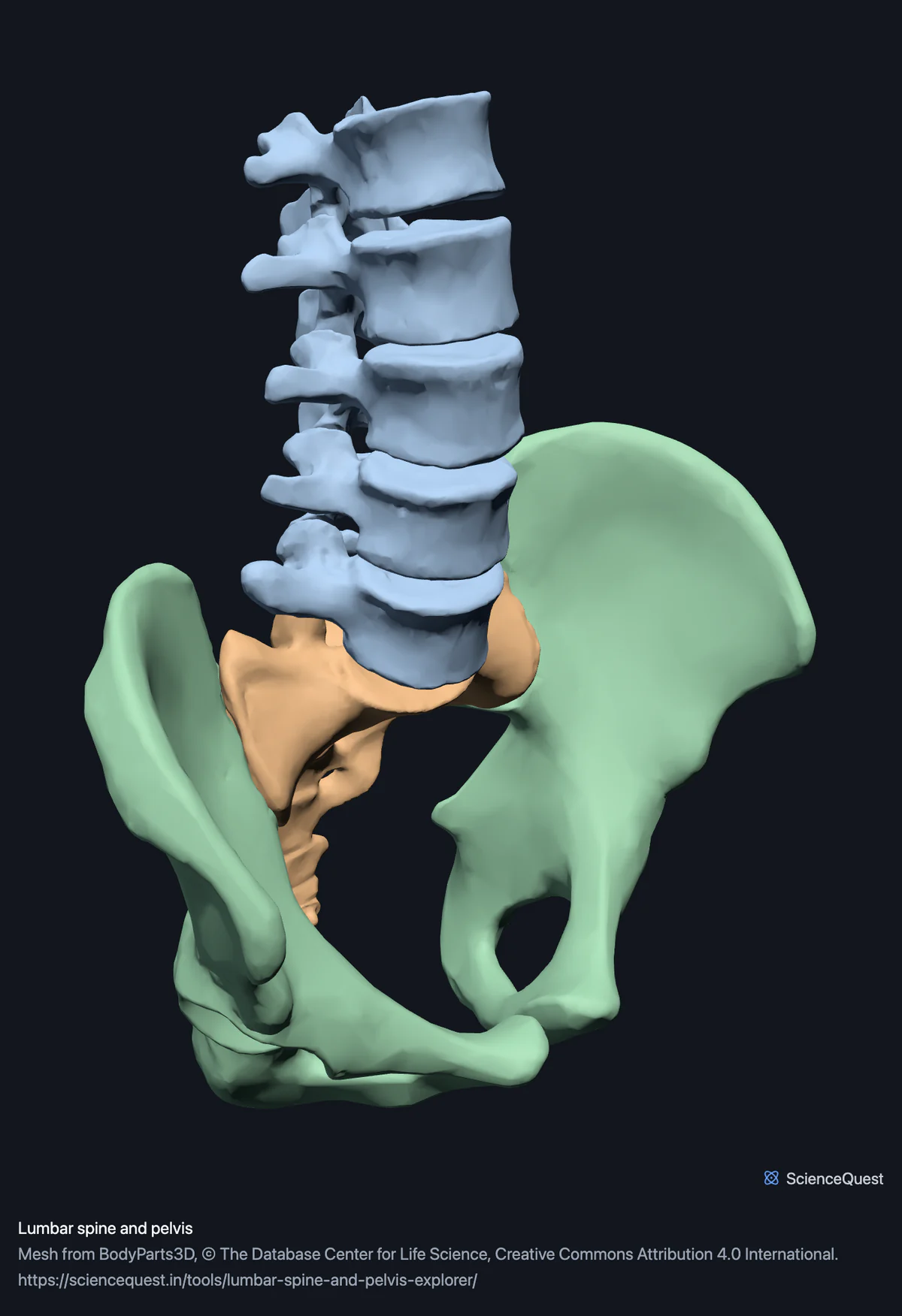
Turn the five lumbar vertebrae, the sacrum and both hip bones in 3D. See the joint that carries the whole upper body from any side.
Visualiser
Lumbar spine and pelvis, 8 bones
Checking this device for a stored copy.
Start here
- Turn to Left or Right and look down the column The vertebral bodies get visibly larger towards the bottom, which is the skeleton answering the load above them.
- Hide the Hip bones group Leaves the column alone, so the lumbosacral junction and the sharp change of direction at it are unobstructed.
- Switch on Cut plane and drag it through the sacrum Shows the fused vertebrae in section.
Bone only. No coccyx, no intervertebral discs, no ligaments and no spinal cord. The discs are what actually transmit load between these bones. The dataset publishes them, but this model leaves them out, so the empty gaps between the vertebral bodies are where they belong; the full skeleton explorer shows them in place.
- 8 bones
- 131k triangles
- 720 KB, on request
- 0.00248 mm worst error per axis
- 5 lumbar vertebrae
- 1 sacrum
- 2 hip bones
Where this comes from
Segmented from imaging of one adult male body, not drawn by an artist, so the shapes are measurements. Two consequences worth knowing: it is one person’s anatomy rather than an average, and faint stepping on some surfaces is the resolution of the original scan rather than a rendering fault.
Meshes from BodyParts3D, © The Database Center for Life Science, under Creative Commons Attribution 4.0 International. Names and identifiers are the dataset’s own: Foundational Model of Anatomy identifiers and English labels, as its parts list gives them. Converted meshes are shared under the same licence.
Changes made: Meshes were converted from binary STL to an indexed mesh with 16-bit quantised vertex positions, and surface normals were recomputed from the geometry. No vertex was moved other than by that quantisation, whose worst-case error on any one coordinate is reported on the page.
Latin terms for 3 of the 8 structures come from English Wikipedia anatomy infoboxes, under Creative Commons Attribution-ShareAlike 4.0, joined by Wikidata property P1402, Foundational Model of Anatomy identifier. Only a term a source states for that structure is used, never one constructed from a family name, which is why the rest carry no Latin rather than a guess.
All 8 bones
Every structure the model contains, group by group. Each name opens the viewer with that structure selected, so a link can point at one bone rather than at the whole region.
Lumbar vertebrae 5
Sacrum 1
- sacrum os sacrum
Hip bones 2
- right hip bone os coxae, os innominatum
- left hip bone os coxae, os innominatum
Citing this tool
Last updated . Add the date you accessed it as well, which a citation of a page that can change asks for. If a specific view matters, cite the page address as it stands in your browser instead: it records what is selected and shown.
Three bones that were once eleven
- Hip bone, one each side
- The ilium, ischium and pubis are separate bones in childhood and fuse at the acetabulum during the teens. The three names survive as names for regions of one bone: the blade, the part you sit on, and the front join.
- Sacrum
- Five vertebrae fused into one wedge. Four transverse ridges across its front surface, each ending at a pair of sacral foramina, show where they were once separate.
- Lumbar vertebrae, five
- The mobile part, and the part that bears the most.
The lordosis is smaller than textbooks draw it
Measured on this specimen, the lumbar vertebrae sit about 18 mm in front of the sacrum once you discount how far above it they are. That is a real curve and a modest one. The exaggerated S in most diagrams leads people to expect something far more dramatic, and it is worth recalibrating against a measurement.
Common questions
Why is the hip bone one bone here rather than three?
Because in an adult it is one bone. The ilium, the ischium and the pubis are three separate bones in childhood, joined by cartilage at the acetabulum, and they fuse during the teens into the single hip bone the dataset segments. The three names survive because they are still the most useful way to refer to regions of it: the ilium is the broad blade, the ischium is what you sit on, and the pubis meets its partner at the front. Turning the model and following the surface from the blade round to the socket is the clearest way to see that it is continuous.
Where is the coccyx?
Not in this dataset. BodyParts3D publishes the coccygeus muscles but no coccyx bone, so the column here ends at the sacrum. That is a genuine gap rather than an editorial choice, and it matters if you are counting: the vertebral column is usually given as 33 vertebrae including three to five fused coccygeal ones, and none of them is here.
Why do L4 and L5 get injured so much more often than the vertebrae above them?
Because they carry the most load and move the most while doing it. Every vertebra supports the weight of everything above it, so the bottom of the lumbar spine carries the most, and it is also where the mobile column meets the fixed sacrum. Look at the model from the side: the vertebral bodies get visibly larger going down, which is the skeleton’s response to that load, and the lumbosacral junction is the sharpest change of direction in the whole column.
What is the lumbar lordosis, and can I see it here?
It is the forward curve of the lower back, and yes, though it is subtler than diagrams suggest. Measured on this specimen the lumbar vertebrae sit only about 18 millimetres in front of the sacrum once you discount how far above it they are. That is a real curve and a small one, which is worth knowing because the exaggerated S drawn in textbooks leads people to expect something much more dramatic. Use the Left or Right view to see it end on.