Eye and Extraocular Muscles 3D Explorer
Peel a real 3D eyeball from sclera and cornea to retina and lens, and see what each of the six eye muscles does and its nerve, in the bones of the orbit.
Visualiser
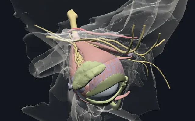
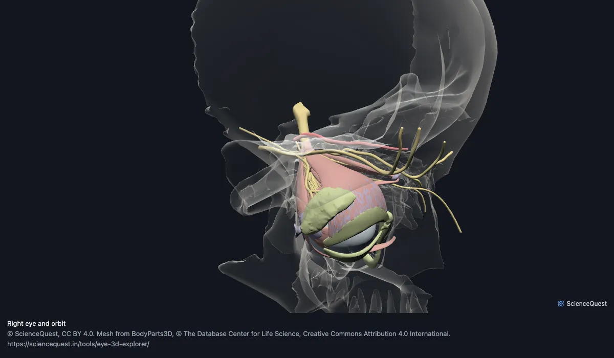
Right eye and orbit, 47 structures
1.1 MB, downloaded only when you press Start exploring and kept on this device afterwards.
Start here
- Back to the eye in its orbit The view the page opens on: the seven bones of the orbit as glass, with the eye, its muscles and its nerves inside them.
- Cut through the eyeball The globe halved from the side, its vitreous body hidden: the sclera, the choroid and the retina lining them, and the lens behind the iris.
- Show the eye muscles from above Bone off, nerves as glass: the recti running back to their ring at the apex of the orbit, and the superior oblique’s tendon in its pulley.
The right eye, its muscles, nerves and tear ducts, with the bones of the orbit. No abducent nerve and no conjunctiva: BodyParts3D 4.0, the release these meshes come from, has no mesh of either. The retina is its optic part only, and the ciliary body its corona ciliaris only, with no orbiculus ciliaris or ciliary muscle. Of the eyelids only the tarsal plates are here, and only the right eye is built, though 4.0 has both. Where the optic nerve’s fibres go after the eye, and the field each lesion cuts, is drawn in the visual field defects explorer.
- 47 structures
- 2 sclera and cornea
- 3 choroid, ciliary body and iris
- 1 retina
- 4 lens, chamber and vitreous
- 7 extraocular muscles
- 5 muscle attachments
- 17 nerves
- 1 ophthalmic artery
- 7 eyelid plates and tear ducts
- 7 bones of the orbit
The frontal bone, sphenoid bone, ethmoid, right maxilla, right zygomatic bone, right lacrimal bone and right palatine bone load with them for context and are not counted above. 54 meshes load in total, and every one of them is listed, nameable and selectable.
Where this comes from
Segmented from MRI of one adult male volunteer, then refined by medical illustrators, who filled in missing detail and sharpened blurred outlines from textbooks and atlases (Mitsuhashi et al., Nucleic Acids Research, 2009). Two consequences worth knowing: it is one person’s anatomy rather than an average, and faint stepping on some surfaces is the resolution of the original scan rather than a rendering fault. These meshes are from release 4.0 (2013), whose release note says it added the internal structure of the eyeball and its appendages and that the data may still contain errors.
Meshes from BodyParts3D, © The Database Center for Life Science, under Creative Commons Attribution 4.0 International. Names and identifiers are the dataset’s own: Foundational Model of Anatomy identifiers and English labels, as its parts list gives them. Converted meshes are shared under the same licence.
Changes made: Meshes were converted from BodyParts3D 4.0’s Wavefront OBJ files, its 99% reduced set, to an indexed mesh with 16-bit quantised vertex positions, and surface normals were recomputed from the geometry. Where the dataset publishes a structure as several element files they were merged into one mesh, except where one element repeats another of the same structure, which was left out; and every piece of a mesh lying across the midline from the side the model shows was removed; the model’s file lists each removal. No vertex was moved other than by that quantisation, whose worst-case error on any one coordinate is reported on the page. On this page it is at most 0.00127 mm per axis. Each model’s file is then compressed without loss, by meshoptimizer’s vertex and index codecs and gzip, and restored exactly before it is drawn. The file holds 261,170 triangles in 1,066 kB, downloaded only when you press Start exploring.
Latin terms for 8 of the 54 structures come from English Wikipedia anatomy infoboxes, under Creative Commons Attribution-ShareAlike 4.0, joined by Wikidata property P1402, Foundational Model of Anatomy identifier. Only a term a source states for that structure is used, never one constructed from a family name, which is why the rest carry no Latin rather than a guess. A few are corrected to the Terminologia Anatomica form where Wikipedia gives an older one, and where several forms are listed, some may still be older synonyms rather than the Terminologia term.
What each eye muscle does, and its nerve
From looking straight ahead. Each name selects the structure in the 3D eye above. Six muscles move the eyeball and the seventh, the levator, lifts the lid; all 7 are in the model, with 16 nerves and nerve branches and the ciliary ganglion.
| Muscle | Moves the eye | Nerve | Arises from |
|---|---|---|---|
| superior rectus | Up (elevation); also in (adduction) and turns the top of the eye toward the nose (intorsion), as it pulls at 23° to the line of sight | Oculomotor nerve (III), upper division | Common tendinous ring (annulus of Zinn) |
| inferior rectus | Down (depression); also in (adduction) and turns the top of the eye away from the nose (extorsion), for the same 23° | Oculomotor nerve (III), lower division | Common tendinous ring (annulus of Zinn) |
| medial rectus | In, toward the nose (adduction) | Oculomotor nerve (III), lower division | Common tendinous ring (annulus of Zinn) |
| lateral rectus | Out, toward the temple (abduction) | Abducent nerve (VI), which supplies no other muscle; BodyParts3D has no mesh of it | Common tendinous ring (annulus of Zinn) |
| superior oblique | Turns the top of the eye toward the nose (intorsion), and moves it down and out; its tendon turns in a pulley, the trochlea, on the frontal bone | Trochlear nerve (IV) | Sphenoid, just above the optic canal, above and medial to the superior rectus’s origin |
| inferior oblique | Turns the top of the eye away from the nose (extorsion); also up (elevation) and out (abduction) | Oculomotor nerve (III), lower division | Orbital surface of the maxilla, just lateral to the nasolacrimal groove, near the front of the orbit’s floor |
| levator palpebrae superioris | Raises the upper eyelid; it does not move the eyeball | Oculomotor nerve (III), upper division | Lesser wing of the sphenoid, above the optic canal |
Sources, for this table and the common questions below: StatPearls (NCBI Bookshelf), the chapters on the extraocular muscles, the eye muscles and each muscle: superior rectus, inferior rectus, medial rectus, lateral rectus, superior oblique and inferior oblique, with cranial nerve 4 for the superior oblique and the orbit for its bones; and Gray’s Anatomy of the Human Body (1918), pages 1005, 1014 to 1015 and 1021 to 1024. Where a chapter lists a muscle’s other actions together, they are given in its order and not ranked.
Parts of the eye and orbit: all 54 named
Every structure the model contains, group by group. Of these, 47 are structures of this region. Each name opens the viewer with that structure selected, so a link can point at one structure rather than at the whole region.
Sclera and cornea 2
- right sclera other tissue
- right cornea other tissue
Choroid, ciliary body and iris 3
- right choroid other tissue
- right corona ciliaris other tissue
- right iris other tissue
Retina 1
- optic part of right retina nervous tissue
Lens, chamber and vitreous 4
- right lens other tissue
- suspensory ligament of right lens other tissue
- anterior chamber of right eyeball other tissue
- right vitreous body other tissue
Extraocular muscles 7
- right superior rectus muscle
- right inferior rectus muscle
- right medial rectus muscle
- right lateral rectus muscle
- right superior oblique muscle
- right inferior oblique muscle
- right levator palpebrae superioris muscle
Muscle attachments 5
- right common tendinous ring other tissue
- trochlea of right superior oblique other tissue
- tendon of right levator palpebrae superioris other tissue
- check ligament of right medial rectus other tissue
- check ligament of right lateral rectus other tissue
Nerves 17
- right optic nerve page nervus opticus nervous tissue
- superior branch of right oculomotor nerve nervous tissue
- inferior branch of right oculomotor nerve nervous tissue
- right trochlear nerve nervous tissue
- right ophthalmic nerve nervous tissue
- right frontal nerve nervous tissue
- right supra-orbital nerve nervous tissue
- right supratrochlear nerve nervous tissue
- right lacrimal nerve nervous tissue
- right nasociliary nerve nervous tissue
- communicating branch of right nasociliary nerve with right ciliary ganglion nervous tissue
- right anterior ethmoidal nerve nervous tissue
- right posterior ethmoidal nerve nervous tissue
- right infratrochlear nerve nervous tissue
- right long ciliary nerve nervous tissue
- right ciliary ganglion nervous tissue
- short ciliary nerve nervous tissue
Ophthalmic artery 1
- right ophthalmic artery blood vessel
Eyelid plates and tear ducts 7
- tarsal plate of right upper eyelid other tissue
- tarsal plate of right lower eyelid other tissue
- right lacrimal gland organ
- right lacrimal lake other tissue
- right lacrimal canaliculus other tissue
- right lacrimal sac other tissue
- right nasolacrimal duct other tissue
Bones of the orbit 7
- frontal bone page os frontale
- sphenoid bone page os sphenoidale
- ethmoid page os ethmoidale
- right maxilla page maxilla
- right zygomatic bone page os zygomaticum, zygoma
- right lacrimal bone page os lacrimale
- right palatine bone page os palatinum
Cite this tool
Citing this tool
Last updated . Add the date you accessed it as well, which a citation of a page that can change asks for. If a specific view matters, cite the page address as it stands in your browser instead: it records what is selected and shown.
On the syllabus: MCAT, 6A: Sensing the environment.
What are the layers of the eye?
Gray’s Anatomy (1918) gives the eyeball three coats, from outside in: a fibrous coat, the sclera behind and the cornea in front; a vascular, pigmented coat, the choroid, ciliary body and iris from back to front; and the retina. The sclera is the opaque back five-sixths of the outer coat and the cornea the clear front sixth. Every one of them is its own mesh here, so hiding the sclera shows the choroid under it, and hiding that shows the optic part of the retina. Inside lie the lens on its suspensory ligament, the anterior chamber in front of the iris and the vitreous body behind the lens.
How six muscles turn one eye
The four recti start at the back of the orbit from one fibrous ring, the common tendinous ring around the optic canal, and run forward to the sclera, each on the side its name gives (Gray 1918). The superior and inferior recti pull at about 23° to the line of sight, so besides lifting or lowering the eye they also turn it in and rotate it (StatPearls). The medial and lateral recti move it purely sideways. The superior oblique runs forward along the upper inner wall to a pulley on the frontal bone, the trochlea, and its tendon turns back to the eyeball behind the equator, which is why it moves the eye down and out; the inferior oblique, which starts near the front of the orbit’s floor, moves it up and out (StatPearls; Gray 1918). Both obliques also rotate the eye about its front to back axis.
An eye examination tests them together: the patient follows a finger tracing a wide letter H, through the nine directions of gaze (StatPearls, Eye Extraocular Muscles). In a palsy of the abducent nerve, for one, the double vision is worst looking toward the side of the weak lateral rectus (StatPearls, Eye Lateral Rectus Muscle).
What BodyParts3D 4.0 adds, and what it leaves out
The meshes are from BodyParts3D release 4.0, whose release note says it added the internal structure of the eyeball and its appendages. Release 3.0, which every other explorer on this site uses, has one mesh for both eyeballs and nothing inside them. 4.0 also moved many parts, so this model is in its own frame: it is never combined with the head, brain or skull models.
Four simplifications come from the data. The abducent nerve is not segmented, so the lateral rectus’s nerve cannot be shown. The retina is its optic part only, the nervous layer that ends at the ora serrata, not the thin layer Gray (1918) traces on over the ciliary processes and the back of the iris. The ciliary body is its corona ciliaris only: Gray (1918, p. 1010) counts the orbiculus ciliaris, the ciliary processes and the ciliary muscle in it, and 4.0 has no mesh of the orbiculus or the muscle. And the long and short ciliary nerves run on into the vitreous body, to about 6 mm from the eyeball’s centre, where Gray (1918, pp. 888 to 889) has them pierce the sclera at the back of the eyeball and run forward on its inner surface; they show inside the globe when its coats are hidden or seen as glass.
What was taken out is listed in the model’s file. The dataset publishes the right optic nerve twice, as two overlapping copies, one of which runs on into the vitreous; this model keeps the one that stops at the back of the eyeball. The short ciliary nerve is one structure in the dataset with a part at each eye, and this model keeps the right eye’s; four right-eye meshes also carry a few stray triangles at the left eye, which were removed. The frontal bone, sphenoid and ethmoid are single midline bones, so they are whole, and the empty left orbit shows the walls the right eye sits in.
Common questions
Which muscles move the eye?
Six: the superior, inferior, medial and lateral recti and the superior and inferior obliques. StatPearls counts seven extraocular muscles, the seventh being the levator palpebrae superioris, which raises the upper eyelid and does not move the eyeball (StatPearls, Eye Extraocular Muscles; Gray 1918). All seven are in this model.
What does the superior oblique do?
It turns the top of the eye toward the nose (intorsion) and moves the eye down and out (StatPearls, Eye Superior Oblique Muscle). Its tendon runs forward through a pulley, the trochlea, on the frontal bone and then back to the upper outer part of the eyeball, behind its equator, so it lifts the back of the eye and the front goes down (StatPearls, Cranial Nerve 4; Gray 1918).
Which nerves supply the eye muscles?
The oculomotor nerve (III) supplies the superior, inferior and medial recti, the inferior oblique and the levator: its upper division the superior rectus and the levator, its lower division the other three. The trochlear nerve (IV) supplies the superior oblique and the abducent nerve (VI) the lateral rectus (StatPearls, Eye Extraocular Muscles; Gray 1918). The model has both oculomotor divisions and the trochlear nerve; BodyParts3D has no abducent nerve.
What are the layers of the eyeball?
Three, from outside in: a fibrous coat, the sclera behind and the cornea in front; a vascular coat, the choroid, ciliary body and iris from back to front; and the retina (Gray 1918). Gray gives the sclera as the back five-sixths of the outer coat and the cornea as the front sixth. Each is a separate mesh here, the ciliary body as its corona ciliaris only, with the lens, its suspensory ligament, the anterior chamber and the vitreous body inside.
Which bones form the orbit?
Seven: the frontal, sphenoid, maxilla, zygomatic, palatine, ethmoid and lacrimal bones (StatPearls, Anatomy, Head and Neck, Orbit). All seven load with the eye, for context, and are not counted in the model’s figure.