Ethmoid
The ethmoid is a single midline bone of the brain case. It makes up much of the upper nasal cavity and the nasal septum, and part of the medial wall of each orbit. (OpenStax A&P 2e, 7.2; StatPearls, Ethmoid Bone)
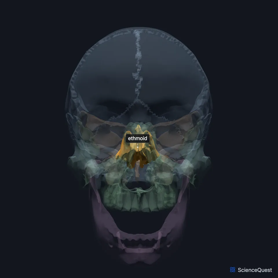
Where is the ethmoid?
Its cribriform plate fills the gap between the frontal bone’s orbital plates in the floor of the anterior cranial fossa, and forms part of the roof of the nasal cavity. (OpenStax A&P 2e, 7.2; Gray 1918, 5d. The Interior of the Skull, 5a. 3. The Frontal Bone and 5a. 6. Ethmoid Bone; StatPearls, Nasal Cavity)
Its side parts lie between each orbit and the upper nasal cavity, so they form the side wall of the nasal cavity and part of the medial wall of the orbit. A plate of the ethmoid called the lamina papyracea separates its air cells from the orbit. (OpenStax A&P 2e, 7.2; StatPearls, Ethmoid Bone)
What are the parts of the ethmoid?
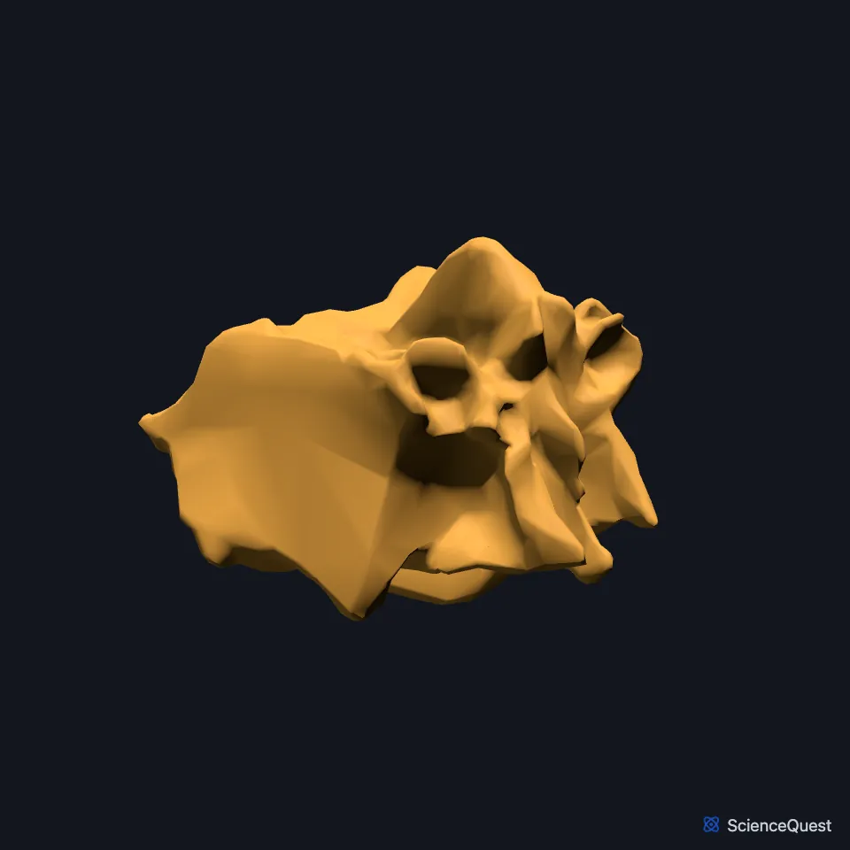
StatPearls lists its parts as the cribriform plate, two ethmoid labyrinths and the perpendicular plate. The crista galli, a small bony projection, rises from the midline. (StatPearls, Ethmoid Bone; OpenStax A&P 2e, 7.2)
The middle and superior nasal conchae, also called turbinates, project from the ethmoid. The inferior nasal concha is a bone of its own. (OpenStax A&P 2e, 7.2; StatPearls, Nasal Concha)
Its side parts, the ethmoid labyrinths, hold the ethmoid air cells, which belong to the paranasal sinuses. A thin plate, the lamina papyracea, separates them from the orbit. (OpenStax A&P 2e, 7.2; StatPearls, Ethmoid Bone and Orbit Bones)
What does the ethmoid do?
The cribriform plate, beside the crista galli, is perforated. The olfactory nerve fibers cross it between the nasal cavity and the olfactory bulb of the brain. (OpenStax A&P 2e, 7.2; Gray 1918, 5a. 6. Ethmoid Bone; StatPearls, Ethmoid Bone and Nasal Cavity)
The crista galli anchors the front end of the falx cerebri, the fold of dura that hangs between the two halves of the brain. (OpenStax A&P 2e, 7.2; StatPearls, Falx Cerebri)
The conchae warm and humidify the air you breathe in and regulate airflow through the nose. StatPearls links the ethmoid air cells with humidification, phonation and ventilation. (OpenStax A&P 2e, 7.2; StatPearls, Nasal Concha and Ethmoid Bone)
Which bones meet the ethmoid?
The septal cartilage forms the front of the nasal septum. Behind it, the perpendicular plate of the ethmoid forms the upper bony part and the vomer the lower part. (OpenStax A&P 2e, 7.2; StatPearls, Nasal Cavity)
StatPearls counts 13 bones that meet the ethmoid: the frontal, the sphenoid, and both nasal bones, maxillae, lacrimal bones, palatine bones and inferior nasal conchae, with the vomer. (StatPearls, Ethmoid Bone)
In the orbit, the ethmoid forms the largest part of the medial wall. The maxilla, the lacrimal bone and a small part of the sphenoid complete it. (OpenStax A&P 2e, 7.2; StatPearls, Orbit Bones; Gray 1918, 5c. The Exterior of the Skull)
The ethmoid also affects drainage of the frontal and maxillary sinuses, through the osteomeatal complex. (StatPearls, Ethmoid Bone)
How does the ethmoid vary?
StatPearls calls the ethmoid sinus the most complex and most variable of the paranasal sinuses. The depth of the olfactory fossa in its roof falls into three Keros types: 1 to 3 mm, 4 to 7 mm and 8 to 16 mm. (StatPearls, Ethmoid Bone)
Where the sources differ
The sources name different parts of the sphenoid in the medial wall of the orbit. StatPearls names the lesser wing, while Gray’s Anatomy (1918) names a small part of the body of the sphenoid, in front of the optic canal. (StatPearls, Orbit Bones; Gray 1918, 5c. The Exterior of the Skull)
| Feature | Detail and source |
|---|---|
| Parts | Cribriform plate, two ethmoid labyrinths, perpendicular plate (StatPearls, Ethmoid Bone) |
| Articulates with | 13 bones: frontal, sphenoid, nasals, maxillae, lacrimals, palatines, inferior nasal conchae, vomer (StatPearls, Ethmoid Bone) |
| Forms | Upper nasal cavity, part of the nasal septum, part of the medial orbital wall (OpenStax A&P 2e, 7.2; StatPearls, Ethmoid Bone) |
Touches in this model
Measured on the BodyParts3D meshes: surfaces within 2 mm of the ethmoid, nearest first.
- Frontal bone in contact
- Left lacrimal bone in contact
- Left maxilla in contact
- Left palatine bone in contact
- Right lacrimal bone in contact
- Right maxilla in contact
- Right palatine bone in contact
- Sphenoid bone in contact
- Vomer in contact
- Eyeball in contact
- Skin in contact
- Right inferior nasal concha 1.9 mm away
Sources
- OpenStax Anatomy and Physiology 2e, section 7.2, The Skull. CC BY-NC-SA 4.0. Read 2026-10-25.
- StatPearls, Anatomy, Head and Neck, Ethmoid Bone. Megan Yu; Shu-Min Wang. Last update July 24, 2023. CC BY-NC-ND 4.0. Read 2026-10-25.
- Gray’s Anatomy of the Human Body, 20th US edition (1918), II. Osteology, 5d. The Interior of the Skull, Upper Surface of the Base of the Skull, and the table of craniometric points (1918 terms: small wings and great wing for lesser and greater wings). Henry Gray. Public domain. Read 2026-10-25.
- Gray’s Anatomy of the Human Body, 20th US edition (1918), II. Osteology, 5a. 3. The Frontal Bone. Henry Gray. Public domain. Read 2026-10-25.
- Gray’s Anatomy of the Human Body, 20th US edition (1918), II. Osteology, 5a. 6. Ethmoid Bone. Henry Gray. Public domain. Read 2026-10-25.
- StatPearls, Anatomy, Head and Neck, Nasal Cavity. John L. Sobiesk; Sunil Munakomi. Last update July 24, 2023. CC BY-NC-ND 4.0. Read 2026-10-25.
- StatPearls, Anatomy, Head and Neck, Nasal Concha. Adegbenro O. Fakoya; Marc H. Hohman; Bianca Georgakopoulos; Patrick H. Le. Last update June 22, 2024. CC BY-NC-ND 4.0. Read 2026-10-25.
- StatPearls, Anatomy, Head and Neck, Orbit Bones. Caleb L. Shumway; Mahsaw Motlagh; Matthew Wade. Last update July 24, 2023. CC BY-NC-ND 4.0. Read 2026-10-25.
- StatPearls, Neuroanatomy, Falx Cerebri. Sunil Munakomi; Felix Jozsa. Last update January 31, 2026. CC BY-NC-ND 4.0. Read 2026-10-25.
- Gray’s Anatomy of the Human Body, 20th US edition (1918), II. Osteology, 5c. The Exterior of the Skull, The Orbits (1918 terms: optic foramen for optic canal). Henry Gray. Public domain. Read 2026-10-25.
The values are facts and free to use; this page’s selection and presentation are © 2026 ScienceQuest.