Eyeball
The adult eyeball is a hollow sphere about 2.5 cm across, set in the bony orbit with only about a sixth of it exposed. Its wall has three layers: the fibrous tunic, the vascular tunic and the retina. (StatPearls, Head and Neck, Eye; OpenStax A&P 2e, 14.1)
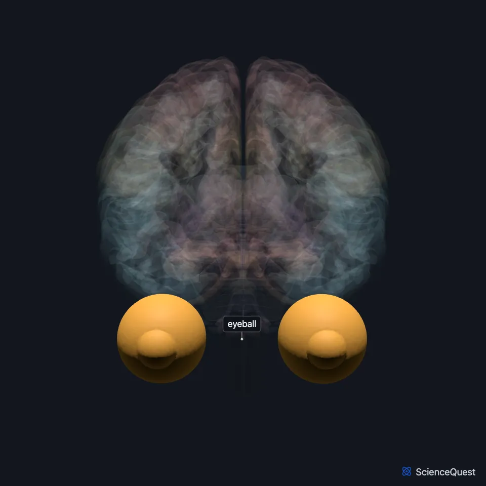
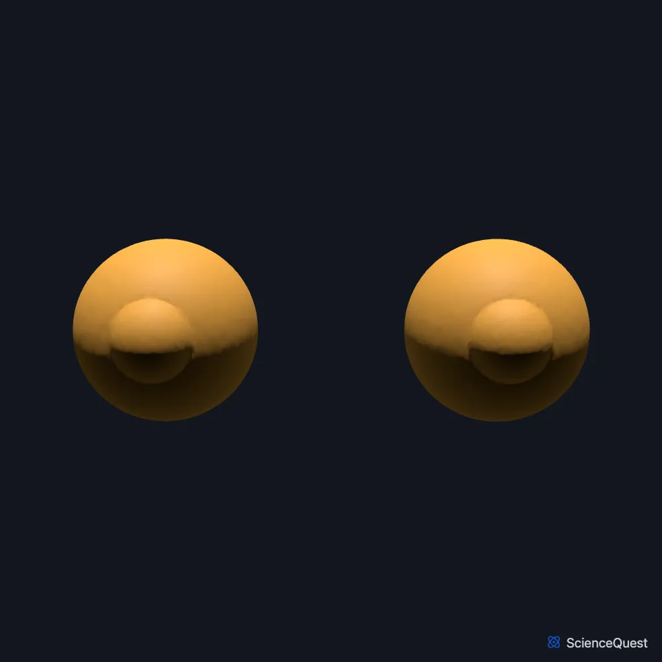
Where is the eyeball?
Each eye sits in a bony orbit of the skull, which shields all but the front sixth of the eyeball. The lacrimal gland, which makes tears, lies in a hollow of the orbital roof, toward the front and outer side. (OpenStax A&P 2e, 14.1; StatPearls, Head and Neck, Eye and Orbit Bones)
What are the layers of the eyeball?
The outer fibrous tunic is the white sclera and the clear cornea. The sclera covers about five sixths of the surface and gives the eyeball its shape, and the cornea covers the front and, with the lens, focuses light onto the retina. (OpenStax A&P 2e, 14.1; StatPearls, Head and Neck, Eye)
The middle vascular tunic, or uvea, is the choroid, the ciliary body and the iris. The choroid’s vessels feed the retina, the ciliary muscle changes the shape of the lens for near and far vision, and the iris sets how much light passes through the pupil. (StatPearls, Head and Neck, Eye)
The inner layer is the retina, with a pigmented layer and a neural layer. Each retina holds about 6 million cones and more than 100 million rods. (StatPearls, Head and Neck, Eye)
The lens divides the inside of the eye into two cavities. The space in front of it holds watery aqueous humor, and the larger space behind it holds the jelly-like vitreous body, which holds the retina against the choroid. (StatPearls, Head and Neck, Eye; OpenStax A&P 2e, 14.1)
How does the eyeball detect light?
Rods and cones respond to light and pass the signal on to the retinal ganglion cells. The axons of about 1.2 million ganglion cells meet at the optic disc and exit the eyeball together as the optic nerve. (OpenStax A&P 2e, 14.1; StatPearls, Cranial Nerve 2 (Optic))
The optic disc has no rods or cones, so it forms a blind spot. The fovea, in the middle of the macula, has only cones and gives the sharpest vision. (StatPearls, Head and Neck, Eye; OpenStax A&P 2e, 14.1)
Cones give color vision and rods let us see in dim light. (StatPearls, Head and Neck, Eye)
What moves and supplies the eyeball?
Six extraocular muscles move the eyeball: the superior, medial, inferior and lateral rectus and the superior and inferior oblique. The four recti start from a fibrous ring around the optic canal at the back of the orbit and attach to the eyeball. (OpenStax A&P 2e, 14.1; StatPearls, Head and Neck, Eye; Gray 1918, 1c. 3. The Accessory Organs of the Eye)
The abducens nerve supplies the lateral rectus, the trochlear nerve the superior oblique, and the oculomotor nerve the other four. (OpenStax A&P 2e, 14.1; StatPearls, Head and Neck: Eye Lateral Rectus Muscle and Head and Neck: Eye Superior Oblique Muscle)
Apart from the eyelids and conjunctiva, the eye is supplied by branches of the internal carotid artery, through the ophthalmic artery. Its sensory nerve is the nasociliary nerve, a branch of the ophthalmic nerve. (StatPearls, Head and Neck, Eye)
Where the sources differ
StatPearls gives visible light as 400 to 700 nm. OpenStax gives 380 to 720 nm. (StatPearls, Head and Neck, Eye; OpenStax A&P 2e, 14.1)
StatPearls’ general eye article pairs the nerves the other way round, giving the lateral rectus to the trochlear nerve and the superior oblique to the abducens. OpenStax and StatPearls’ own articles on the lateral rectus and the superior oblique give the lateral rectus to the abducens and the superior oblique to the trochlear nerve. (StatPearls, Head and Neck, Eye, Head and Neck: Eye Lateral Rectus Muscle and Head and Neck: Eye Superior Oblique Muscle; OpenStax A&P 2e, 14.1)
| Feature | Detail and source |
|---|---|
| Diameter | About 2.5 cm in an adult (StatPearls, Head and Neck, Eye) |
| Layers | Fibrous tunic, vascular tunic (uvea), retina (StatPearls, Head and Neck, Eye) |
| Photoreceptors | About 6 million cones and over 100 million rods per retina (StatPearls, Head and Neck, Eye) |
| Leaves the eye | Optic nerve, at the optic disc (OpenStax A&P 2e, 14.1; StatPearls, Cranial Nerve 2 (Optic)) |
Touches in this model
Measured on the BodyParts3D meshes: surfaces within 2 mm of the eyeball, nearest first, 12 of 18.
- Left optic nerve in contact
- Right optic nerve in contact
- Ethmoid in contact
- Left zygomatic bone in contact
- Orbital part of left orbicularis oculi in contact
- Orbital part of right orbicularis oculi in contact
- Palpebral part of left orbicularis oculi in contact
- Palpebral part of right orbicularis oculi in contact
- Right lacrimal bone in contact
- Right zygomatic bone in contact
- Skin in contact
- Sphenoid bone in contact
Sources
- StatPearls, Anatomy, Head and Neck, Eye. Ibraheem Rehman; Bita Hazhirkarzar; Bhupendra C. Patel. Last update July 24, 2023. CC BY-NC-ND 4.0. Read 2026-10-25.
- OpenStax Anatomy and Physiology 2e, section 14.1, Sensory Perception. CC BY-NC-SA 4.0. Read 2026-10-25.
- StatPearls, Anatomy, Head and Neck, Orbit Bones. Caleb L. Shumway; Mahsaw Motlagh; Matthew Wade. Last update July 24, 2023. CC BY-NC-ND 4.0. Read 2026-10-25.
- StatPearls, Neuroanatomy, Cranial Nerve 2 (Optic). Austen M. Smith; Craig N. Czyz. Last update November 7, 2022. CC BY-NC-ND 4.0. Read 2026-10-25.
- Gray’s Anatomy of the Human Body, 20th US edition (1918), X. The Organs of the Senses and the Common Integument, 1c. 3. The Accessory Organs of the Eye (1918 terms: optic foramen for optic canal, bulb of the eye for eyeball). Henry Gray. Public domain. Read 2026-10-25.
- StatPearls, Anatomy, Head and Neck: Eye Lateral Rectus Muscle. Alejandro Fernández Cabrera; Juan Suárez-Quintanilla. Last update August 8, 2023. CC BY-NC-ND 4.0. Read 2026-10-25.
- StatPearls, Anatomy, Head and Neck: Eye Superior Oblique Muscle. Ahmed Abdelhady; Bhupendra C. Patel; Sanah Aslam; Daifallah M. Al Aboud. Last update August 7, 2023. CC BY-NC-ND 4.0. Read 2026-10-25.
The values are facts and free to use; this page’s selection and presentation are © 2026 ScienceQuest.