Optic nerve
The optic nerve is the second cranial nerve (CN II). It carries visual information from the retina and is purely sensory. (StatPearls, Cranial Nerve 2 (Optic); OpenStax A&P 2e, 13.4)
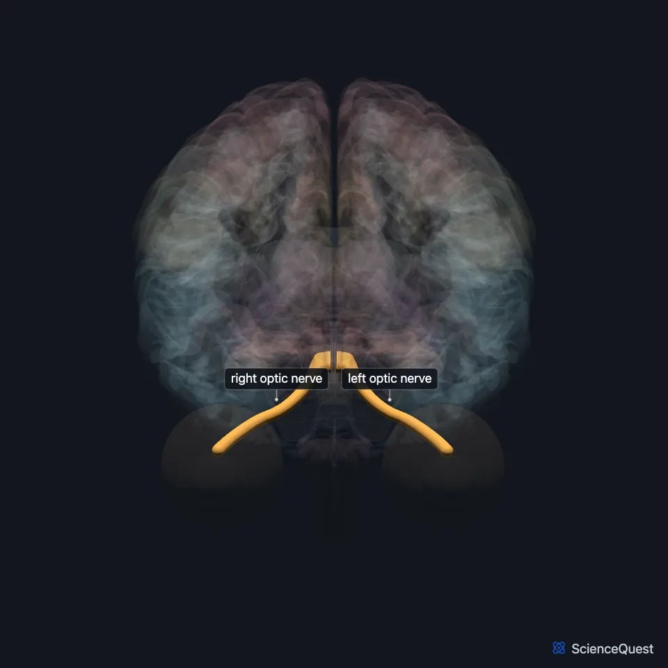
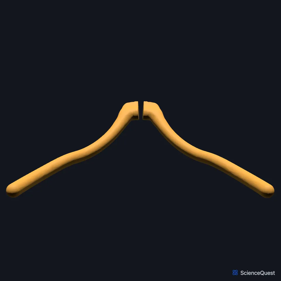
Where does the optic nerve run?
In the adult, the axons of about 1.2 million retinal ganglion cells gather at the optic disc to form it. They leave the eyeball through openings in the sclera called the lamina cribrosa. (StatPearls, Cranial Nerve 2 (Optic))
With the ophthalmic artery, it leaves the orbit through the optic canal and reaches the middle cranial fossa. There the two optic nerves meet at the optic chiasm and continue as the optic tracts. (StatPearls, Cranial Nerve 2 (Optic))
What does the optic nerve do?
At the chiasm, fibers from the nasal half of each retina cross to the opposite optic tract, while fibers from the temporal half stay on the same side. So each optic tract carries the opposite half of the visual field. (StatPearls, Cranial Nerve 2 (Optic); OpenStax A&P 2e, 14.2)
Most optic tract fibers end in the lateral geniculate nucleus of the thalamus, which projects to the visual cortex in the occipital lobe. Other optic tract fibers go to the pretectal nucleus of the midbrain, for the pupillary light reflex, and to the superior colliculus. A very small number of fibers leave at the chiasm, before the tract forms, and go to the suprachiasmatic nucleus of the hypothalamus, which controls the circadian rhythm. (OpenStax A&P 2e, 14.2; StatPearls, Cranial Nerve 2 (Optic))
It is the afferent limb of the pupillary light reflex and of the accommodation reflex. Light shone into one eye makes both pupils constrict. (StatPearls, Cranial Nerve 2 (Optic))
What is the optic nerve made of?
It grows out of the diencephalon and belongs to the central nervous system. Oligodendrocytes myelinate it, and it is sheathed in all three meningeal layers, which are continuous with those of the brain. (StatPearls, Cranial Nerve 2 (Optic))
The ganglion cell axons stay unmyelinated until they pass the lamina cribrosa, so no myelin lies in the path of light inside the retina. (StatPearls, Cranial Nerve 2 (Optic))
Axons from each quarter of the retina keep together in the matching quarter of the nerve. Inside the skull, the fibers from the macula move to the center of the nerve. (StatPearls, Optic Chiasm)
Because its sheaths are continuous with the meninges, raised pressure inside the skull can spread along the nerve and show as papilledema on fundoscopy. (StatPearls, Cranial Nerve 2 (Optic))
What lies next to the optic nerve?
The optic chiasm, where the nerves end, sits at the base of the brain below the hypothalamus and about 10 mm above the pituitary gland. (StatPearls, Optic Chiasm)
Its main artery is the ophthalmic artery, the first branch of the internal carotid artery, which runs below and to the outer side of the nerve in the optic canal. The central retinal artery pierces the nerve about 12 mm behind the eyeball and runs forward to the inner retina. (StatPearls, Cranial Nerve 2 (Optic))
Where the sources differ
OpenStax counts the superior colliculus among the three major targets of the optic tract. StatPearls calls the fibers that reach it another small group. (OpenStax A&P 2e, 14.2; StatPearls, Cranial Nerve 2 (Optic))
| Feature | Detail and source |
|---|---|
| Cranial nerve | II, purely sensory (StatPearls, Cranial Nerve 2 (Optic)) |
| Axons | About 1.2 million, from retinal ganglion cells (StatPearls, Cranial Nerve 2 (Optic)) |
| Leaves the orbit through | Optic canal (StatPearls, Cranial Nerve 2 (Optic)) |
| Ends at | Optic chiasm (StatPearls, Cranial Nerve 2 (Optic)) |
| Main artery | Ophthalmic artery (StatPearls, Cranial Nerve 2 (Optic)) |
Touches in this model
Measured on the BodyParts3D meshes: surfaces within 2 mm of the optic nerve, nearest first.
- Eyeball in contact
- Hypothalamus in contact
- Lamina terminalis in contact
- Left optic tract in contact
- Optic chiasm in contact
- Orbital gyri and straight gyri, both sides in contact
- Right optic tract in contact
- Skin in contact
- Sphenoid bone in contact
- Third ventricle 0.7 mm away
- Tuber cinereum 0.8 mm away
- Pituitary gland 1.7 mm away
Sources
- StatPearls, Neuroanatomy, Cranial Nerve 2 (Optic). Austen M. Smith; Craig N. Czyz. Last update November 7, 2022. CC BY-NC-ND 4.0. Read 2026-10-25.
- OpenStax Anatomy and Physiology 2e, section 13.4, The Peripheral Nervous System. CC BY-NC-SA 4.0. Read 2026-10-25.
- OpenStax Anatomy and Physiology 2e, section 14.2, Central Processing. CC BY-NC-SA 4.0. Read 2026-10-25.
- StatPearls, Neuroanatomy, Optic Chiasm. Ashley C. Ireland; Iverson B. Carter. Last update May 1, 2023. CC BY-NC-ND 4.0. Read 2026-10-25.
The values are facts and free to use; this page’s selection and presentation are © 2026 ScienceQuest.