Optic chiasm
The optic chiasm is where the two optic nerves meet and some of their fibers cross the midline. It is named for this X shape, after the Greek letter chi. (StatPearls, Optic Chiasm; OpenStax A&P 2e, 16.3)
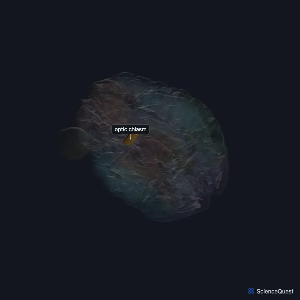
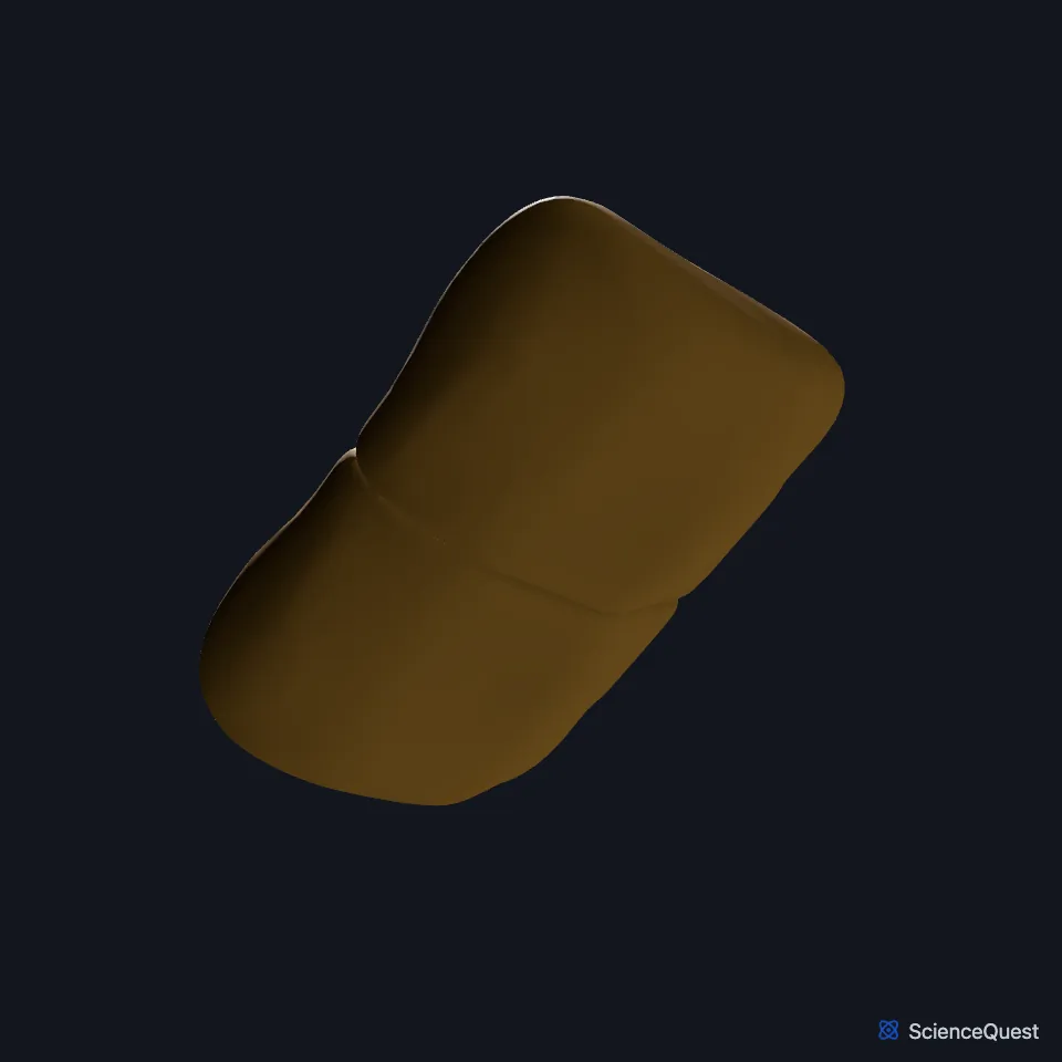
Where is the optic chiasm?
It lies at the base of the brain, under the hypothalamus, in the suprasellar cistern. (StatPearls, Optic Chiasm)
It forms part of the front of the floor of the third ventricle, and the anterior communicating and anterior cerebral arteries lie close to it. (StatPearls, Optic Chiasm)
Which fibers cross at the optic chiasm?
Axons from the medial, or nasal, half of each retina cross here; axons from the lateral half stay on their own side. (OpenStax A&P 2e, 14.2; StatPearls, Optic Chiasm)
Because of this crossing, each optic tract, and so each cerebral hemisphere, carries the opposite half of the visual field: the right tract carries the left visual field. (OpenStax A&P 2e, 14.2; StatPearls, Optic Chiasm and Visual Pathway)
The crossing fibers come from the medial retinae, so they carry the outer, peripheral part of the visual field. (OpenStax A&P 2e, 16.3; StatPearls, Optic Chiasm)
Inside the chiasm, crossing fibers from the upper nasal retina run toward the back and those from the lower nasal retina toward the front. (StatPearls, Optic Chiasm)
What connects to the optic chiasm?
The optic nerves enter the skull through the optic canals and meet here. Behind it, the regrouped fibers continue as the optic tracts. (OpenStax A&P 2e, 16.3 and 14.2; StatPearls, Optic Chiasm and Cranial Nerve 2 (Optic))
Most optic tract fibers end in the lateral geniculate nucleus of the thalamus, and others reach the superior colliculus of the midbrain. A small set of retinal axons, the retinohypothalamic tract, reaches the suprachiasmatic nucleus of the hypothalamus. (OpenStax A&P 2e, 14.2; StatPearls, Optic Chiasm, Visual Pathway and Nucleus Suprachiasmatic)
The retina began as part of the embryonic diencephalon: in the third week of gestation, optic grooves grow out from it and form the optic vesicles. (OpenStax A&P 2e, 13.1 and 14.2; StatPearls, Visual Pathway)
The pituitary gland lies about 10 mm below. A pituitary growth can press on the crossing fibers and take away the outer, temporal half of the visual field in both eyes. (OpenStax A&P 2e, 14.2 and 16.3; StatPearls, Optic Chiasm)
Where the sources differ
The sources disagree on where the pituitary sits. OpenStax A&P 2e puts it directly below the chiasm; StatPearls puts the chiasm in front of and above the gland, about 10 mm above the pituitary fossa. (OpenStax A&P 2e, 16.3; StatPearls, Pituitary Gland and Optic Chiasm)
The sources give the field loss from chiasm compression different names. OpenStax A&P 2e calls it bilateral hemianopia; StatPearls calls it bitemporal hemianopsia, loss of the temporal visual field in both eyes from compression of the crossing nasal fibers. (OpenStax A&P 2e, 14.2; StatPearls, Optic Chiasm)
Touches in this model
Measured on the BodyParts3D meshes: surfaces within 2 mm of the optic chiasm, nearest first, 12 of 15.
- Hypothalamus in contact
- Lamina terminalis in contact
- Left optic nerve in contact
- Left optic tract in contact
- Right optic nerve in contact
- Right optic tract in contact
- Third ventricle in contact
- Tuber cinereum in contact
- Orbital gyri and straight gyri, both sides 0.1 mm away
- Right stria terminalis 0.2 mm away
- Left stria terminalis 0.5 mm away
- Peduncle of midbrain 1.7 mm away
Sources
- StatPearls, Neuroanatomy, Optic Chiasm. Ashley C. Ireland; Iverson B. Carter. Last update May 1, 2023. CC BY-NC-ND 4.0. Read 2026-10-25.
- OpenStax Anatomy and Physiology 2e, section 16.3, The Cranial Nerve Exam. CC BY-NC-SA 4.0. Read 2026-10-25.
- OpenStax Anatomy and Physiology 2e, section 14.2, Central Processing. CC BY-NC-SA 4.0. Read 2026-10-25.
- StatPearls, Neuroanatomy, Visual Pathway. Mohit Gupta; Ashley C. Ireland; Adekunle E. Omole; Bruno Bordoni. Last update March 23, 2026. CC BY-NC-ND 4.0. Read 2026-10-25.
- StatPearls, Neuroanatomy, Cranial Nerve 2 (Optic). Austen M. Smith; Craig N. Czyz. Last update November 7, 2022. CC BY-NC-ND 4.0. Read 2026-10-25.
- StatPearls, Neuroanatomy, Nucleus Suprachiasmatic. Joe M. Das; Marjorie V. Launico. Last update September 14, 2026. CC BY-NC-ND 4.0. Read 2026-10-25.
- OpenStax Anatomy and Physiology 2e, section 13.1, The Embryologic Perspective. CC BY-NC-SA 4.0. Read 2026-10-25.
- StatPearls, Anatomy, Head and Neck, Pituitary Gland. Muthu Kuzhali Ganapathy; Prasanna Tadi. Last update July 24, 2023. CC BY-NC-ND 4.0. Read 2026-10-25.
The values are facts and free to use; this page’s selection and presentation are © 2026 ScienceQuest.