Third ventricle
The third ventricle is the narrow midline cavity between the left and right halves of the diencephalon. (OpenStax A&P 2e, 13.3; StatPearls, Ventricular System)
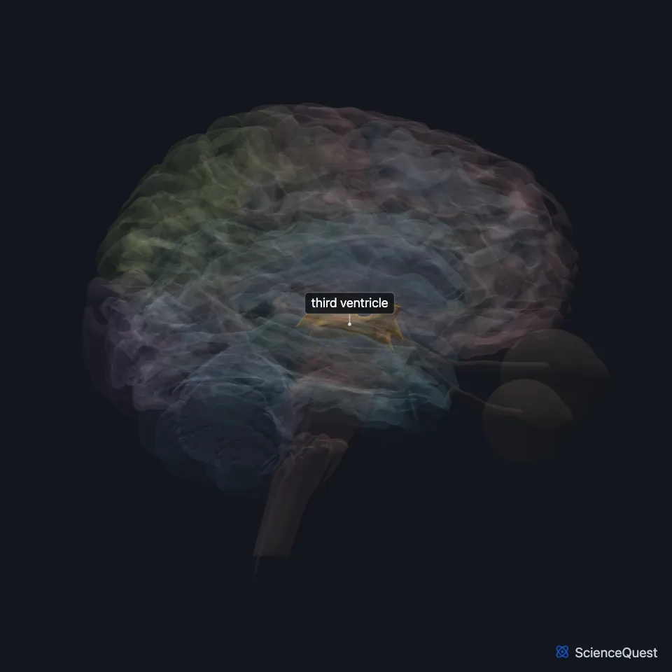
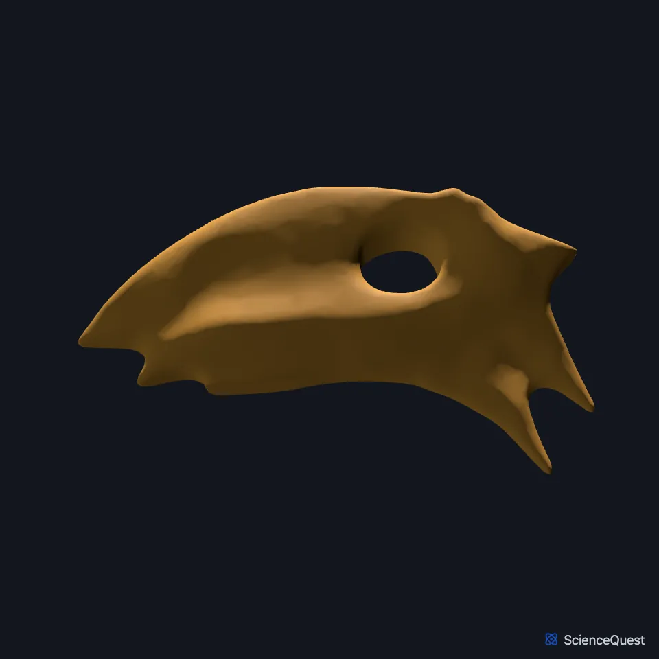
Where is the third ventricle?
The medial walls of the thalamus and hypothalamus bound it. A curved hypothalamic sulcus divides each side wall into a thalamic upper part and a hypothalamic lower part. (OpenStax A&P 2e, 13.3; StatPearls, Ventricular System)
The interthalamic adhesion, or massa intermedia, crosses it to join the two thalami. StatPearls notes it may be missing in about 30% of brains. (OpenStax A&P 2e, 13.3; StatPearls, Ventricular System)
From front to back, its floor is the optic chiasm, the tuber cinereum and infundibulum, the mammillary body, the posterior perforated substance and the tegmentum of the midbrain. (StatPearls, Ventricular System)
What forms the walls of the third ventricle?
Its front wall, from top to bottom, is the columns of the fornix, the anterior commissure and the lamina terminalis. Its back wall is the pineal body, the posterior commissure and the opening of the cerebral aqueduct. (StatPearls, Ventricular System)
Its roof is a sheet of ependyma under a fold of pia mater, the tela choroidea, from which two fringes hang down as the choroid plexus of the third ventricle. (StatPearls, Ventricular System)
It pushes out into five recesses: infundibular, optic, anterior, pineal and suprapineal. The infundibular recess reaches down into the stalk of the pituitary gland. (StatPearls, Ventricular System)
What does the third ventricle do?
It passes CSF from the lateral ventricles, which open into it through the interventricular foramina, on to the cerebral aqueduct, and more CSF is made in it on the way. (OpenStax A&P 2e, 13.3; StatPearls, Ventricular System and Choroid Plexus)
Like the rest of the ventricular system, it develops from the single hollow of the embryo’s neural tube. (OpenStax A&P 2e, 13.1 and 13.3; StatPearls, Ventricular System)
Where does fluid leave the third ventricle?
The cerebral aqueduct leaves it at the back, runs through the midbrain and opens into the fourth ventricle. (OpenStax A&P 2e, 13.3; StatPearls, Cerebral Aqueduct (Sylvian) and Ventricular System)
The aqueduct, about 18 mm long, is the narrowest part of the ventricular system and the most common site of a block. (StatPearls, Ventricular System)
Touches in this model
Measured on the BodyParts3D meshes: surfaces within 2 mm of the third ventricle, nearest first, 12 of 32.
- Anterior commissure in contact
- Cerebral aqueduct in contact
- Choroid plexus of left cerebral hemisphere in contact
- Choroid plexus of right cerebral hemisphere in contact
- Corpus callosum in contact
- Habenula in contact
- Hypothalamus in contact
- Interventricular foramen in contact
- Lamina terminalis in contact
- Left fornix of forebrain in contact
- Left stria medullaris of thalamus in contact
- Left thalamus in contact
Sources
- OpenStax Anatomy and Physiology 2e, section 13.3, Circulation and the Central Nervous System. CC BY-NC-SA 4.0. Read 2026-10-25.
- StatPearls, Neuroanatomy, Ventricular System. Saraswati Satyanarayan Shenoy; Forshing Lui. Last update July 24, 2023. CC BY-NC-ND 4.0. Read 2026-10-25.
- StatPearls, Neuroanatomy, Choroid Plexus. Kinaan Javed; Vamsi Reddy; Forshing Lui. Last update July 24, 2023. CC BY-NC-ND 4.0. Read 2026-10-25.
- OpenStax Anatomy and Physiology 2e, section 13.1, The Embryologic Perspective. CC BY-NC-SA 4.0. Read 2026-10-25.
- StatPearls, Neuroanatomy, Cerebral Aqueduct (Sylvian). Jessica M. Rubino; Jeffery P. Hogg. Last update July 24, 2023. CC BY-NC-ND 4.0. Read 2026-10-25.
The values are facts and free to use; this page’s selection and presentation are © 2026 ScienceQuest.