Lateral ventricle
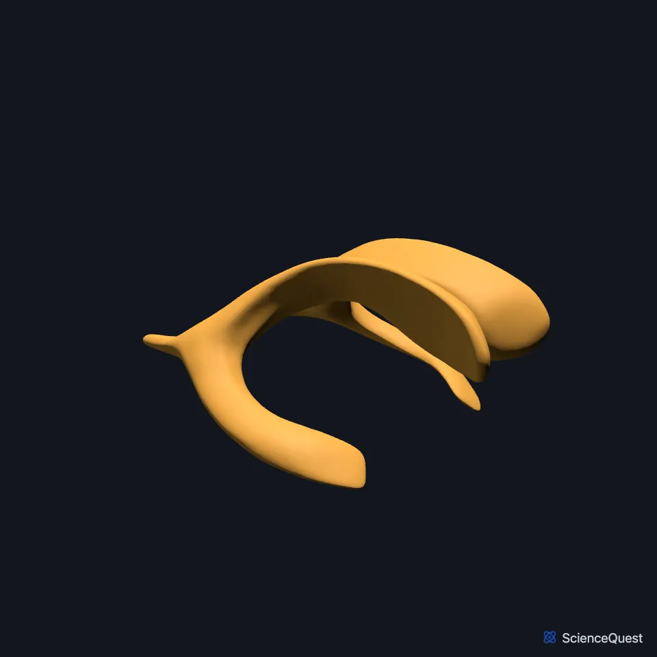
The lateral ventricles are two C-shaped cavities, one in each cerebral hemisphere, lined by ependyma and filled with cerebrospinal fluid (CSF). (StatPearls, Ventricular System)
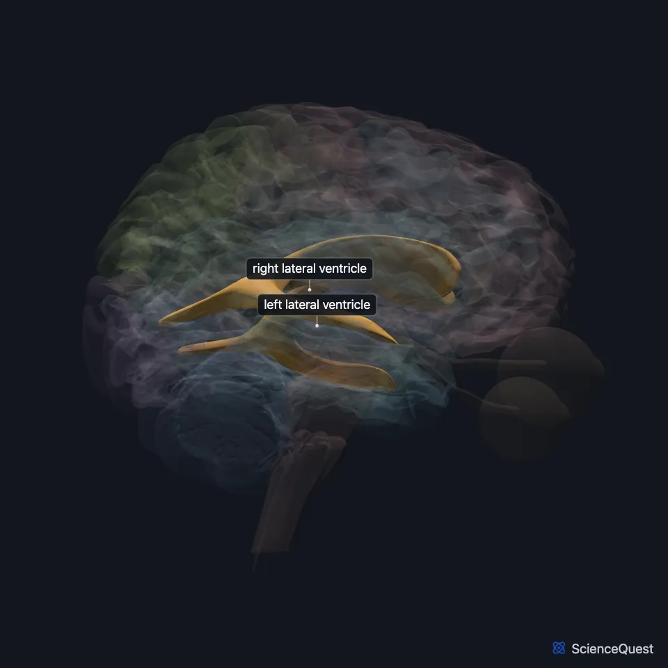
Where are the lateral ventricles?
They lie deep in the cerebrum and are the largest of the four ventricles. (OpenStax A&P 2e, 13.3; StatPearls, Fourth Ventricle)
A thin vertical sheet of nervous tissue, the septum pellucidum, separates the two. (StatPearls, Ventricular System)
Each one curves in a C, around the back of the thalamus and then down and forward into the temporal lobe. (OpenStax A&P 2e, 13.3; StatPearls, Ventricular System)
What are the parts of a lateral ventricle?
Each has a central part, the body, and three horns: anterior, posterior and inferior. Each has a capacity of 7 to 10 ml. (StatPearls, Ventricular System)
The body lies in the parietal lobe, from the interventricular foramen back to the splenium of the corpus callosum. The anterior horn reaches into the frontal lobe, the posterior horn into the occipital lobe, and the inferior horn, the largest, curves around the back of the thalamus into the temporal lobe. (StatPearls, Ventricular System)
What forms the walls of a lateral ventricle?
The caudate nucleus lines it: the head forms its lateral wall, the body lies beside its body, and the tail lies above its temporal horn. (StatPearls, Nucleus Caudate)
The corpus callosum forms the roof of the body, and the floor of the body includes part of the upper surface of the thalamus. (StatPearls, Ventricular System)
What does the lateral ventricle do?
Its choroid plexus makes most of the CSF, with help from the ependymal lining of the ventricles. The lateral ventricles make the most CSF because of their larger surface area. (OpenStax A&P 2e, 13.3; StatPearls, Fourth Ventricle)
CSF carries metabolic waste out of the nervous tissue and cushions the brain and spinal cord. All the plexuses together make about 500 milliliters a day. (OpenStax A&P 2e, 13.3; StatPearls, Choroid Plexus and Cerebrospinal Fluid)
Where does fluid from the lateral ventricles go?
CSF leaves each one through an interventricular foramen into the third ventricle; the foramina join the front of the lateral ventricles to the third ventricle. (OpenStax A&P 2e, 13.3; StatPearls, Ventricular System)
Touches in this model
Measured on the BodyParts3D meshes: surfaces within 2 mm of the lateral ventricle, nearest first, 12 of 35.
- Choroid plexus of left cerebral hemisphere in contact
- Choroid plexus of right cerebral hemisphere in contact
- Commissure of fornix of forebrain in contact
- Corpus callosum in contact
- Interventricular foramen in contact
- Left amygdala in contact
- Left caudate nucleus in contact
- Left cingulate gyrus in contact
- Left fornix of forebrain in contact
- Left fusiform gyrus in contact
- Left hippocampus in contact
- Left occipital lobe in contact
Sources
- StatPearls, Neuroanatomy, Ventricular System. Saraswati Satyanarayan Shenoy; Forshing Lui. Last update July 24, 2023. CC BY-NC-ND 4.0. Read 2026-10-25.
- OpenStax Anatomy and Physiology 2e, section 13.3, Circulation and the Central Nervous System. CC BY-NC-SA 4.0. Read 2026-10-25.
- StatPearls, Neuroanatomy, Fourth Ventricle. Zachary K. Roesch; Prasanna Tadi. Last update August 7, 2023. CC BY-NC-ND 4.0. Read 2026-10-25.
- StatPearls, Neuroanatomy, Nucleus Caudate. Margaret E. Driscoll; Pradeep C. Bollu; Prasanna Tadi. Last update July 24, 2023. CC BY-NC-ND 4.0. Read 2026-10-25.
- StatPearls, Neuroanatomy, Choroid Plexus. Kinaan Javed; Vamsi Reddy; Forshing Lui. Last update July 24, 2023. CC BY-NC-ND 4.0. Read 2026-10-25.
- StatPearls, Neuroanatomy, Cerebrospinal Fluid. Konstantinos Margetis; Luke J. Weisbrod; Marjorie V. Launico. Last update July 7, 2025. CC BY-NC-ND 4.0. Read 2026-10-25.
The values are facts and free to use; this page’s selection and presentation are © 2026 ScienceQuest.