Caudate nucleus
The caudate nucleus is one of the basal nuclei deep in the cerebrum: a long, C-shaped nucleus, one in each hemisphere. (OpenStax A&P 2e, 13.2; StatPearls, Nucleus Caudate)
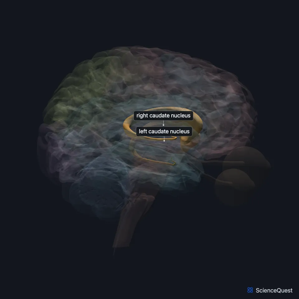
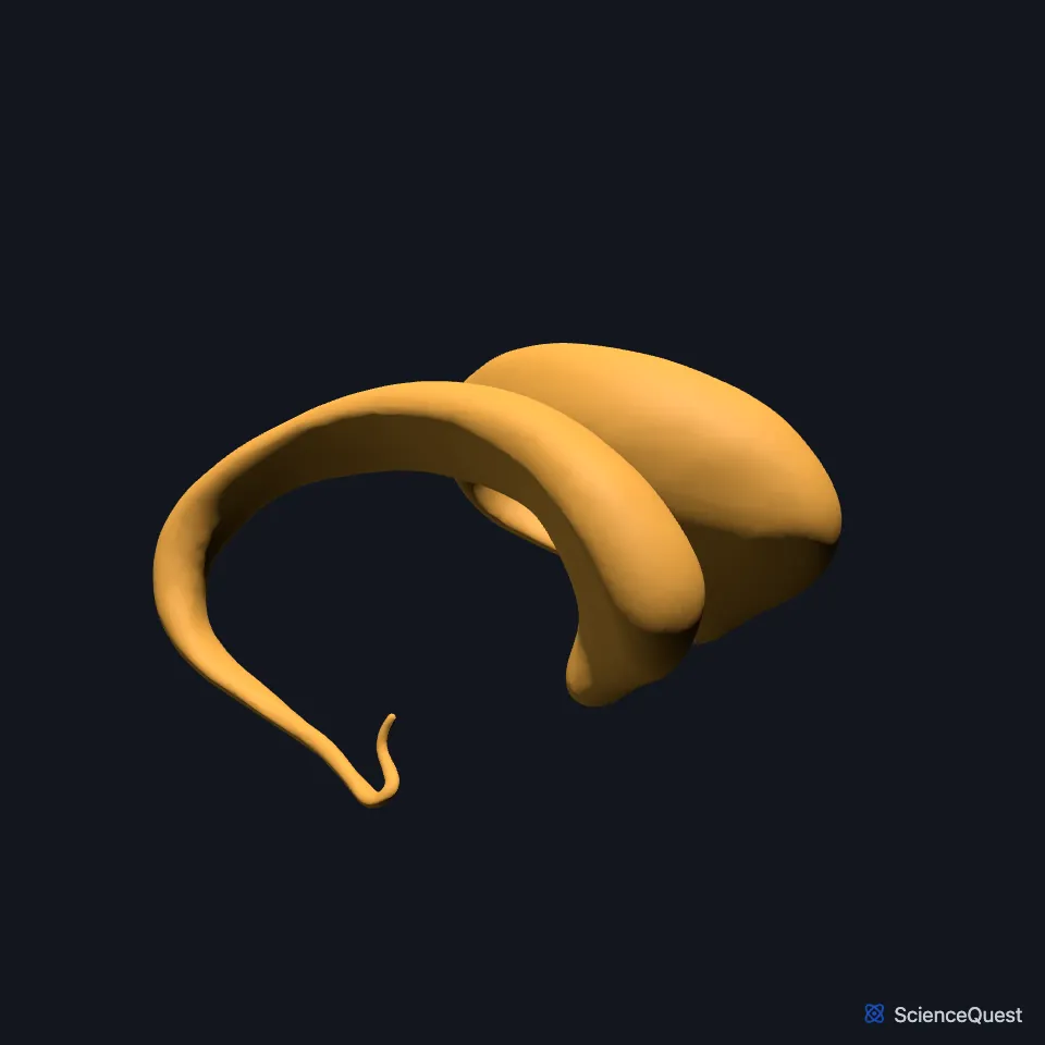
Where is the caudate nucleus?
It curves in a C along the lateral ventricle. Its head forms the side wall of the ventricle, its body runs beside the body of the ventricle, and its tail lies above the temporal horn. (OpenStax A&P 2e, 13.2; StatPearls, Nucleus Caudate and Ventricular System)
It hugs the lateral ventricle: its head forms the lateral wall of the ventricle, its body lies beside the body of the ventricle, and its tail lies above the temporal horn. (StatPearls, Nucleus Caudate)
The two caudate nuclei lie near the thalamus in the center of the brain, and the head of each meets the thalamus at its front outer edge. (StatPearls, Nucleus Caudate and Thalamus)
What does the caudate nucleus do?
With the putamen it forms the striatum, which receives the input that the cerebral cortex sends to the basal nuclei. (OpenStax A&P 2e, 13.2; StatPearls, Putamen and Basal Ganglia)
The basal nuclei weigh what the cortex intends against input from deep brain stem nuclei, and so make a movement more or less likely. The direct pathway releases movement and the indirect pathway holds it back. (OpenStax A&P 2e, 13.2; StatPearls, Basal Ganglia)
Beyond planning movement, it works in learning, memory, reward, motivation and emotion. (StatPearls, Nucleus Caudate)
Most of its neurons are medium spiny neurons that use GABA and inhibit other parts of the basal ganglia. (StatPearls, Nucleus Caudate)
What are the parts of the caudate nucleus?
Each has a large head at the front, a body, and a thin tail that curves forward again, so one coronal slice can cut both the head and the tail. (StatPearls, Nucleus Caudate)
The front part connects with the lateral and medial prefrontal cortex and works in working memory and executive function. The tail works with the inferior temporal lobe on visual processing and movement control. (StatPearls, Nucleus Caudate)
What connects to the caudate nucleus?
Its input comes from the cortex, mostly the frontal lobe on the same side. Its output, like the rest of the striatum’s, goes to the globus pallidus and the substantia nigra. The internal segment of the globus pallidus and the substantia nigra pars reticulata then project to the thalamus. (StatPearls, Nucleus Caudate, Putamen and Basal Ganglia; OpenStax A&P 2e, 13.2)
The internal capsule, a large white matter tract, separates it from the putamen. This model shows the anterior limb of the internal capsule, which runs between the head of the caudate nucleus and the lentiform nucleus. The corticospinal tract descends further back, in the posterior limb, between the thalamus and the lentiform nucleus. (OpenStax A&P 2e, 13.2; StatPearls, Internal Capsule)
Dopamine from the substantia nigra pars compacta, in the midbrain, reaches the striatum and switches it between two pathways: it activates the direct pathway and inhibits the indirect one. (OpenStax A&P 2e, 13.2; OpenStax Psychology 2e, 3.4; StatPearls, Substantia Nigra and Basal Ganglia)
Where the sources differ
The sources differ on where the caudate nucleus sends its output. StatPearls Neuroanatomy, Nucleus Caudate lists the hippocampus, globus pallidus and thalamus; OpenStax A&P 2e and StatPearls Neuroanatomy, Putamen send the striatum’s output to the globus pallidus and the substantia nigra. (StatPearls, Nucleus Caudate and Putamen; OpenStax A&P 2e, 13.2)
Touches in this model
Measured on the BodyParts3D meshes: surfaces within 2 mm of the caudate nucleus, nearest first, 12 of 25.
- Anterior limb of left internal capsule in contact
- Anterior limb of right internal capsule in contact
- Choroid plexus of left cerebral hemisphere in contact
- Choroid plexus of right cerebral hemisphere in contact
- Corpus callosum in contact
- Interventricular foramen in contact
- Left amygdala in contact
- Left fornix of forebrain in contact
- Left lateral ventricle in contact
- Left putamen in contact
- Left stria terminalis in contact
- Left thalamus in contact
Sources
- OpenStax Anatomy and Physiology 2e, section 13.2, The Central Nervous System. CC BY-NC-SA 4.0. Read 2026-10-25.
- StatPearls, Neuroanatomy, Nucleus Caudate. Margaret E. Driscoll; Pradeep C. Bollu; Prasanna Tadi. Last update July 24, 2023. CC BY-NC-ND 4.0. Read 2026-10-25.
- StatPearls, Neuroanatomy, Ventricular System. Saraswati Satyanarayan Shenoy; Forshing Lui. Last update July 24, 2023. CC BY-NC-ND 4.0. Read 2026-10-25.
- StatPearls, Neuroanatomy, Thalamus. Tyler J. Torrico; Sunil Munakomi. Last update July 24, 2023. CC BY-NC-ND 4.0. Read 2026-10-25.
- StatPearls, Neuroanatomy, Putamen. Mehrnoosh Ghandili; Sunil Munakomi. Last update January 30, 2023. CC BY-NC-ND 4.0. Read 2026-10-25.
- StatPearls, Neuroanatomy, Basal Ganglia. Carly B. Young; Vamsi Reddy; James Sonne. Last update July 24, 2023. CC BY-NC-ND 4.0. Read 2026-10-25.
- StatPearls, Neuroanatomy, Internal Capsule. Marc Christopher Emos; Mahammed Z. Khan Suheb; Sanjeev Agarwal. Last update June 5, 2023. CC BY-NC-ND 4.0. Read 2026-10-25.
- OpenStax Psychology 2e, section 3.4, The Brain and Spinal Cord. CC BY-NC-SA 4.0. Read 2026-10-25.
- StatPearls, Neuroanatomy, Substantia Nigra. James Sonne; Vamsi Reddy; Morris R. Beato. Last update September 10, 2024. CC BY-NC-ND 4.0. Read 2026-10-25.
The values are facts and free to use; this page’s selection and presentation are © 2026 ScienceQuest.