Hippocampus
The hippocampus is a three-layered structure in the temporal lobe, named for its resemblance to a seahorse. (OpenStax Behavioral Neuroscience, 18.3; Neuroscience Online, Limbic System: Hippocampus)
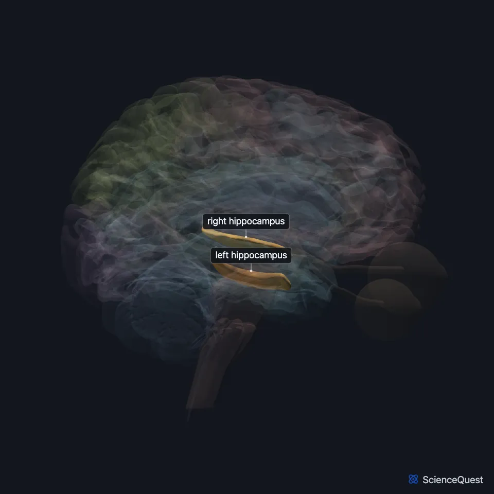
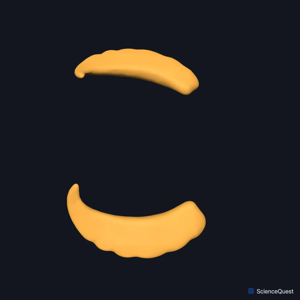
Where is the hippocampus?
Each temporal lobe holds one, alongside the inferior horn of the lateral ventricle. (Neuroscience Online, Limbic System: Hippocampus; StatPearls, Hippocampus)
The amygdala abuts its front end; you expose it by peeling away the uncus. (Neuroscience Online, Limbic System: Amygdala)
Rodent and primate hippocampi are named differently along their long axis. The rodent dorsal hippocampus corresponds to the posterior hippocampus in primates, and the rodent ventral part to the anterior part. (OpenStax Behavioral Neuroscience, 18.3; Strange et al. 2014, Nat Rev Neurosci; Fanselow and Dong 2010, Neuron)
How is the hippocampus built?
The hippocampus and dentate gyrus are cortex with three layers, against six in the neocortex. They are inside-out: the molecular layer lies on the inside and the polymorphic layer on the outside. (Neuroscience Online, Limbic System: Hippocampus)
A major route through it runs one way: from the entorhinal cortex to the dentate gyrus by the perforant pathway, to region CA3 by the mossy fibers, then to CA1 by the Schaffer collaterals. (Neuroscience Online, Limbic System: Hippocampus)
What does the hippocampus do?
It sits in the medial temporal lobe, its front end at the amygdala. The two work in long-term memory: the hippocampus with memories of facts and events, the amygdala with emotional memories. (OpenStax A&P 2e, 13.2; StatPearls, Hippocampus; Neuroscience Online, Learning and Memory)
It is a major part of the brain’s system for declarative memory, the conscious memory for facts and events. (Neuroscience Online, Learning and Memory)
After surgery for epilepsy, the patient H.M. could not form new memories for facts and events, yet he could still recall events from the distant past. He could still learn new skills, without knowing he had them. (Neuroscience Online, Learning and Memory; OpenStax Psychology 2e, 8.2; OpenStax Behavioral Neuroscience, 18.1)
How does the hippocampus connect to the rest of the brain?
Its two main routes in and out are the fornix and the entorhinal cortex, and its inputs and outputs travel bundled together in the same paths. (Neuroscience Online, Limbic System: Hippocampus)
Through the fornix it reaches the septal nuclei, the anterior nucleus of the thalamus and the mammillary bodies. (Neuroscience Online, Limbic System: Hippocampus)
The two hippocampi communicate through the hippocampal commissure and through fornix fibers that cross in the anterior commissure. (Neuroscience Online, Limbic System: Hippocampus)
Where the sources differ
Sources disagree on whether the hippocampal formation includes the entorhinal cortex. Neuroscience Online lists the dentate gyrus, the hippocampus proper and the subicular cortex. OpenStax Behavioral Neuroscience lists the hippocampus proper, the dentate gyrus, the subiculum, presubiculum and parasubiculum, and the entorhinal cortex. (Neuroscience Online, Limbic System: Hippocampus; OpenStax Behavioral Neuroscience, 18.3)
Sources describe H.M.’s surgery differently. OpenStax Psychology 2e section 3.4 says his hippocampus and amygdala were removed, section 8.2 says both temporal lobes (hippocampi), and Neuroscience Online says his hippocampus, on both sides. OpenStax Behavioral Neuroscience 18.1 says the anterior two-thirds of both temporal lobes, a bilateral temporal lobectomy. (OpenStax Psychology 2e, 3.4 and 8.2; Neuroscience Online, Learning and Memory; OpenStax Behavioral Neuroscience, 18.1)
Sources differ on how many of H.M.’s older memories survived. Neuroscience Online says he had all of his memories from before the operation, and OpenStax Psychology 2e 8.2 says he could still remember events from before the surgery. OpenStax Behavioral Neuroscience 18.1 says he could recall events from the distant past, which suggests that remote and recent memories may be stored in different brain regions. (Neuroscience Online, Learning and Memory; OpenStax Psychology 2e, 8.2; OpenStax Behavioral Neuroscience, 18.1)
| Feature | Detail and source |
|---|---|
| Lobe | Temporal, beside the inferior horn of the lateral ventricle (Neuroscience Online, Limbic System: Hippocampus; StatPearls, Hippocampus) |
| Cortical layers | 3 (Neuroscience Online, Limbic System: Hippocampus) |
| Main cortical input | Entorhinal cortex (OpenStax Behavioral Neuroscience, 18.3; Neuroscience Online, Limbic System: Hippocampus) |
| Main tract | Fornix (Neuroscience Online, Limbic System: Hippocampus) |
Touches in this model
Measured on the BodyParts3D meshes: surfaces within 2 mm of the hippocampus, nearest first, 12 of 26.
- Choroid plexus of left cerebral hemisphere in contact
- Choroid plexus of right cerebral hemisphere in contact
- Commissure of fornix of forebrain in contact
- Left amygdala in contact
- Left fornix of forebrain in contact
- Left fusiform gyrus in contact
- Left inferior temporal gyrus in contact
- Left lateral geniculate body in contact
- Left lateral ventricle in contact
- Left parahippocampal gyrus in contact
- Left thalamus in contact
- Right amygdala in contact
Sources
- OpenStax Introduction to Behavioral Neuroscience, section 18.3, Explicit Memories: Episodic and Semantic Memories. CC BY-NC-SA 4.0. Read 2026-10-25.
- Neuroscience Online, McGovern Medical School, Section 4, Chapter 5, Limbic System: Hippocampus. Anthony Wright, Ph.D.. Last update Revised 07 July 2026. Open access; no reuse licence stated. Read 2026-10-25.
- StatPearls, Neuroanatomy, Hippocampus. Leslie A. Fogwe; Vamsi Reddy; Fassil B. Mesfin. Last update July 20, 2023. CC BY-NC-ND 4.0. Read 2026-10-25.
- Neuroscience Online, McGovern Medical School, Section 4, Chapter 6, Limbic System: Amygdala. Anthony Wright, Ph.D.. Last update Revised 20 July 2026. Open access; no reuse licence stated. Read 2026-10-25.
- Nature Reviews Neuroscience, Functional organization of the hippocampal longitudinal axis, Nat Rev Neurosci 15, pages 655 to 669 (abstract, PMID 25234264). Strange BA, Witter MP, Lein ES, Moser EI. Abstract read on Europe PMC; no reuse licence stated. Read 2026-10-25.
- Neuron, Are the dorsal and ventral hippocampus functionally distinct structures?, Neuron 65, pages 7 to 19 (author manuscript on PMC, PMC2822727; PMID 20152109). Fanselow MS, Dong HW. Author manuscript on PMC; no reuse licence stated. Read 2026-10-25.
- OpenStax Anatomy and Physiology 2e, section 13.2, The Central Nervous System. CC BY-NC-SA 4.0. Read 2026-10-25.
- Neuroscience Online, McGovern Medical School, Section 4, Chapter 7, Learning and Memory. John H. Byrne, Ph.D.. Last update Reviewed 06 July 2026. Open access; no reuse licence stated. Read 2026-10-25.
- OpenStax Psychology 2e, section 8.2, Parts of the Brain Involved with Memory. CC BY-NC-SA 4.0. Read 2026-10-25.
- OpenStax Introduction to Behavioral Neuroscience, section 18.1, Memory is Classified Based on Time Course and Type of Information Stored. CC BY-NC-SA 4.0. Read 2026-10-25.
- OpenStax Psychology 2e, section 3.4, The Brain and Spinal Cord. CC BY-NC-SA 4.0. Read 2026-10-25.
The values are facts and free to use; this page’s selection and presentation are © 2026 ScienceQuest.