Heart and Lung Auscultation Points Explorer
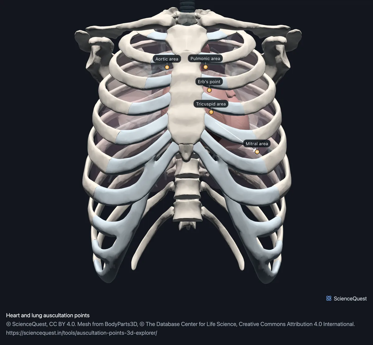
See the cardiac auscultation areas and the lung sites on real 3D ribs over the heart and lungs, with what is heard best at each and the source for it.
Visualiser
Heart: five areas.
Five areas: four named for a valve, and Erb’s point
- The diaphragm picks up high-pitched sounds and the bell low-pitched ones. Press the bell lightly: pressed hard, the skin acts as a diaphragm and the low sounds are lost. Merck Manual, Cardiac Auscultation
- The order differs between sources. Open RN usually starts at the aortic area and moves through the pulmonic area, Erb’s point and the tricuspid area to the mitral area, with the diaphragm and then the bell. Merck typically starts over the apex with the patient on the left side, then has the patient roll supine and works up the left sternal border space by space and down from the right upper sternal border, and also listens over the left axilla and above the clavicles. The patient then sits up for the back, and leans forward for aortic and pulmonic diastolic murmurs. Open RN Nursing Skills, 9.3; Merck Manual, Cardiac Auscultation
Pick an area to see what to listen for there and, for the four valve areas, where that valve lies.
Start here
- Heart The five cardiac areas in the order of APE To Man; pick one for what to listen for there, with its source.
- Lungs 14 sites front, sides and back, each with the lobe beneath it; a pin hides while its side faces away.
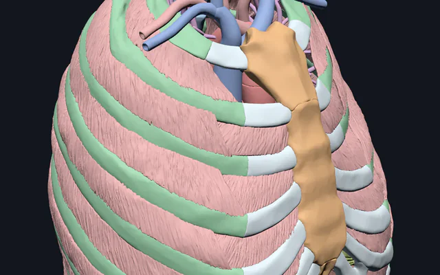
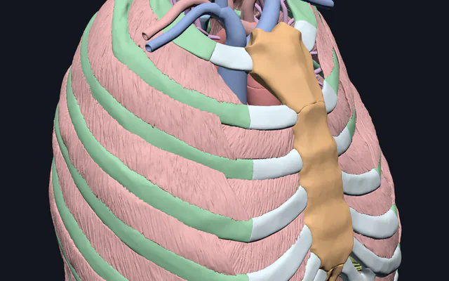
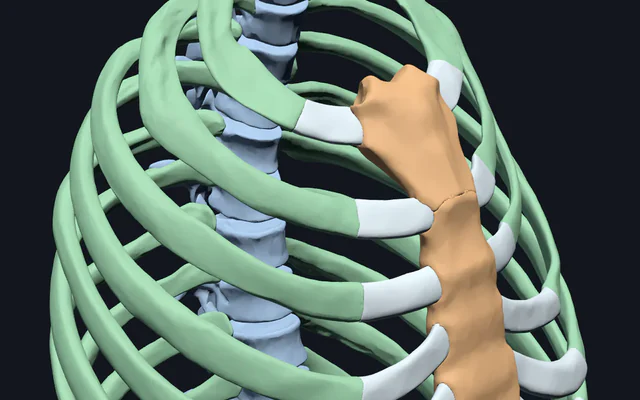
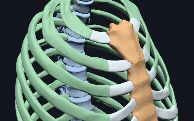
- Click a bone Every rib, cartilage and vertebra is named, so you can count down from the second rib at the sternal angle (OpenStax Anatomy and Physiology 2e, 7.4).
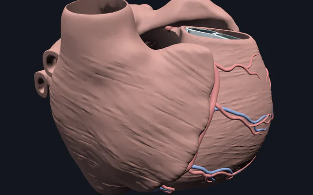
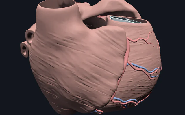
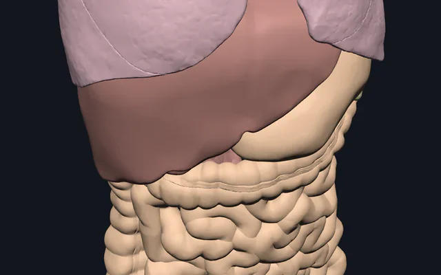
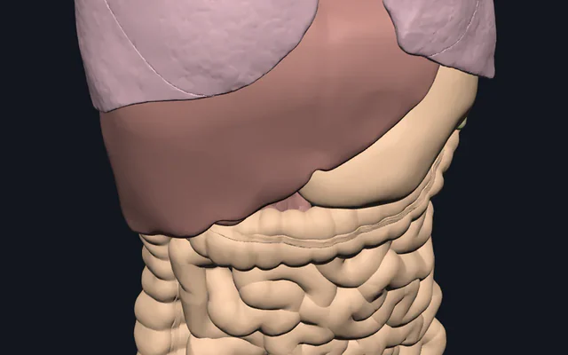
Bone and costal cartilage from the BodyParts3D skeleton, the heart wall and three of its valves, the five lung lobes with the trachea and bronchi, and the ascending aorta and its arch. No skin, breast tissue, muscle or fat, so each pin sits on the surface of the ribs rather than the skin; the two apex sites, above the clavicles, sit on the lung itself. No aortic valve: BodyParts3D 3.0, the release these meshes come from, has no aortic valve mesh, so its place is the root of the ascending aorta. No pulmonary trunk or pulmonary artery either, so the pulmonic area’s arrow starts at the pulmonary valve.
Where this comes from
Segmented from MRI of one adult male volunteer, then refined by medical illustrators, who filled in missing detail and sharpened blurred outlines from textbooks and atlases (Mitsuhashi et al., Nucleic Acids Research, 2009). Two consequences worth knowing: it is one person’s anatomy rather than an average, and faint stepping on some surfaces is the resolution of the original scan rather than a rendering fault.
Meshes from BodyParts3D, © The Database Center for Life Science, under Creative Commons Attribution 4.0 International. Names and identifiers are the dataset’s own: Foundational Model of Anatomy identifiers and English labels, as its parts list gives them. Converted meshes are shared under the same licence.
Changes made: Meshes were converted from binary STL to an indexed mesh with 16-bit quantised vertex positions, and surface normals were recomputed from the geometry. No vertex was moved other than by that quantisation, whose worst-case error on any one coordinate is reported on the page. On this page it is at most 0.00311 mm. The model is a ScienceQuest selection of 72 meshes from four BodyParts3D models, simplified from 1.99 million to 0.69 million triangles with no surface moved more than 0.63 mm. The pins are ScienceQuest’s measurement of these meshes. Each model’s file is then compressed without loss, by meshoptimizer’s vertex and index codecs and gzip, and restored exactly before it is drawn.
Latin terms for 12 of the 72 structures come from English Wikipedia anatomy infoboxes, under Creative Commons Attribution-ShareAlike 4.0, joined by Wikidata property P1402, Foundational Model of Anatomy identifier. Only a term a source states for that structure is used, never one constructed from a family name, which is why the rest carry no Latin rather than a guess. A few are corrected to the Terminologia Anatomica form where Wikipedia gives an older one, and where several forms are listed, some may still be older synonyms rather than the Terminologia term.
Cite this tool
Citing this tool
Last updated . Add the date you accessed it as well, which a citation of a page that can change asks for. If a specific view matters, cite the page address as it stands in your browser instead: it records what is selected and shown.
What are the five cardiac auscultation points?
Five places on the chest, in APE To Man order: the aortic area at the second right intercostal space beside the sternum, the pulmonic area at the second left, Erb’s point at the third left, the tricuspid area at the fourth left and the mitral area at the fifth left, on the midclavicular line (Open RN Nursing Skills, 9.3).
Finding the spaces on the model
Each pin was placed by measuring this model’s own cartilages. The manubrium meets the body of the sternum at the sternal angle, where the second rib attaches, which makes it the landmark for counting the ribs below (OpenStax Anatomy and Physiology 2e, 7.4), and each space takes the number of the rib above it (Radiopaedia, Intercostal spaces). On this model the middle of the second costal cartilage meets the sternum within 0.1 in (3 mm) of it. Measured on vertical lines 0.2 in (5 mm) outside the sternum’s left edge, where the pins stand, the second intercostal space is 0.6 in (14 mm) tall, the third 0.4 in (10 mm), the fourth 0.3 in (8 mm) and the fifth 0.2 in (4 mm), each shorter than the one above. The midclavicular lines, drawn in Open RN Nursing Skills, 10.3, Figure 10.2, are taken here as the vertical through the midpoint of each clavicle’s two ends, and are 6.3 in (160 mm) apart.
The heart wall’s apex lies 0.4 in (11 mm) inside the left midclavicular line, behind the fifth costal cartilage on its own vertical, between the fourth and fifth spaces, so the mitral area, which Open RN puts in the fifth space on the line itself, sits just below and lateral to it.
The areas are not over their valves
The pins do not lie over the valves they are named for. Measured here, the pulmonic area is 1.8 in (46 mm) from the pulmonary valve, the aortic area 3.0 in (76 mm) from the root of the ascending aorta, the tricuspid area 3.0 in (75 mm) from the tricuspid valve and the mitral area 4.5 in (115 mm) from the mitral valve, which lies 3.9 in (98 mm) deeper in the chest than its pin.
Lung sites and the lobes beneath them
The 14 lung sites are ScienceQuest’s own sampling of the fields, not a set any source gives: Open RN’s Figures 10.5 and 10.6 show 12 sites on the front and 12 on the back, side to side, the first pair above the clavicles, and name no rib spaces. In this body the apices above the clavicles and the second spaces on the midclavicular lines lie over the upper lobes, the right fourth space over the right middle lobe and the left fourth over the left upper lobe. The sixth spaces on the sides lie over the right lower lobe and the left lower lobe, and on the back the sites at the third thoracic spine lie over the upper lobes, those at the sixth over the right lower lobe and the left lower lobe. So in this model the lower lobes are reached from the back and the sides, and the right middle lobe from the front.
Where the sources differ
- Aortic regurgitation. The Merck Manual places its early diastolic murmur loudest at the third or fourth left parasternal space or at the apex, which is Erb’s point and below; Open RN lists it under the aortic and pulmonic areas.
- The order to listen in. Open RN usually starts at the aortic area and works through to the mitral area, with the diaphragm and then the bell. Merck typically starts over the apex with the patient on the left side, then has the patient roll supine, works up the left sternal border and down from the right upper sternal border, and also listens over the left axilla and above the clavicles, then with the patient sitting up and leaning forward.
- The tricuspid area. Open RN places it at the fourth space and gives the fourth and fifth for its murmurs; Merck gives the left middle or lower sternal border, or the epigastrium, for tricuspid regurgitation.
- The apex. Open RN puts the mitral (apical) area at the fifth space on the midclavicular line. Merck feels the apical impulse between the fourth and fifth spaces, just medial to that line, which is where this model’s apex lies.
What the model leaves out
- Skin, muscle, fat and breast tissue. The pins sit on the ribs, so on a patient each area is a little further from the heart than it is here.
- The aortic valve and the pulmonary trunk. BodyParts3D 3.0, the release these meshes come from, has no aortic valve mesh (its 4.0 parts list adds one, FMA7236), so the root of the ascending aorta stands for it. Release 3.0 has no separate pulmonary trunk either, and its one pulmonary artery mesh (FMA66326) is not in this model, so that pin’s card names only the lung.
- Breathing. The lungs are one static shape from one scan, so the lobe under a site is where this scan put it.
- One body. The meshes are one adult man’s, so every distance here is his.
Sources
- Ernstmeyer K, Christman E, editors. Nursing Skills, 2nd edition, chapter 9.3, Cardiovascular Assessment. Open RN, WisTech Open, 2023.
- Ernstmeyer K, Christman E, editors. Nursing Skills, 2nd edition, chapter 10.3, Respiratory Assessment. Open RN, WisTech Open, 2023.
- Gupta JI, Shea MJ. Cardiac Auscultation. Merck Manual Professional Version, full review April 2025.
- Armstrong GP. Aortic Stenosis. Merck Manual Professional Version, full review June 2025.
- Armstrong GP. Aortic Regurgitation. Merck Manual Professional Version, full review June 2025.
- Armstrong GP. Mitral Regurgitation. Merck Manual Professional Version, full review June 2025.
- Armstrong GP. Mitral Stenosis. Merck Manual Professional Version, full review June 2025.
- Armstrong GP. Tricuspid Regurgitation. Merck Manual Professional Version, full review June 2025.
- Armstrong GP. Pulmonary Stenosis. Merck Manual Professional Version, full review June 2025.
- Dezube R. Evaluation of the Patient With Pulmonary Issues. Merck Manual Professional Version, full review May 2025.
- Gupta JI, Shea MJ. Cardiovascular Examination. Merck Manual Professional Version, full review April 2025.
- Betts JG, Young KA, Wise JA, et al. Anatomy and Physiology 2e, section 7.4, The Thoracic Cage. OpenStax, 2022.
- Smith S, Silverstone L, et al. Intercostal spaces. Reference article, Radiopaedia.org, rID 37903, revised September 2024.
- BodyParts3D, release 3.0 (20110915), English parts list. Database Center for Life Science, 2011.
- BodyParts3D, release 4.0 (20130619), English part-of parts list. Database Center for Life Science, 2013.
Common questions
Where are the aortic, pulmonic, tricuspid and mitral areas?
The aortic area, the second intercostal space at the right sternal border; the pulmonic area, the second space at the left sternal border; Erb’s point, the third space at the left sternal border; the tricuspid area, the fourth space at the left sternal border; and the mitral area, the fifth space at the left midclavicular line (Open RN Nursing Skills, 2023); Merck feels the apical impulse between the fourth and fifth spaces, just medial to that line.
Where is Erb’s point, and what is heard there?
At the third intercostal space on the left sternal border, directly below the pulmonic area (Open RN). Merck places the early diastolic murmur of aortic regurgitation loudest at the third or fourth left parasternal space or at the apex; Open RN lists that murmur under the aortic and pulmonic areas instead.
Is each auscultation area over its valve?
No. Measured on this model, the pulmonic area’s pin is 1.8 in (46 mm) from the pulmonary valve, the aortic area’s 3.0 in (76 mm) from the root of the ascending aorta, the tricuspid area’s 3.0 in (75 mm) from the tricuspid valve and the mitral area’s 4.5 in (115 mm) from the mitral valve. The arrows in the explorer run from each valve to its area.
Where do you listen to the lungs?
Over every field of the chest, including the flanks and the front (Merck). Open RN listens side to side, comparing each site with its mirror, with the diaphragm on skin rather than clothing and not over the scapulae, the clavicles or the breasts; its figures show 12 sites on the front and 12 on the back, the first pair above the clavicles. Vesicular sounds are the normal sound over most lung fields (Merck), and bronchial sounds are heard over the trachea and larynx (Open RN).