Back and Spine Explorer
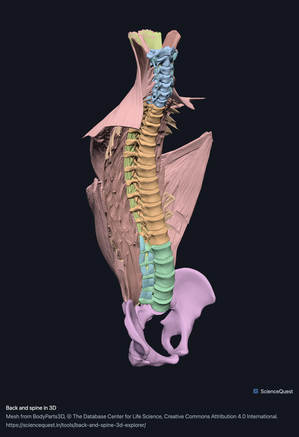
The whole vertebral column with the back muscles over it, in layers: 85 structures, peeled superficial to deep the way a dissection goes.
Visualiser
Vertebral column and pelvis, 27 bones
Checking this device for a stored copy.
Start here
- Hide the Superficial layer band Fourteen meshes off in one press: trapezius in three parts a side, latissimus dorsi, levator scapulae and rhomboid major and minor. That is the first cut of any back dissection.
- Press Label on Erector spinae Selects the whole band, so a saved figure names all sixteen. Up to 24 structures can be labelled at once, which is why a band this size works and a larger one is capped.
- Filter to Bone and open Thoracic The twelve thoracic vertebrae alone, in order, with the muscles still loaded and one press away.
Bone and muscle. No spinal cord and no nerve roots: BodyParts3D segments only the central canal of the cord, a thin tube, and files that with the brain. No intervertebral discs either, because they belong to the full skeleton model rather than to the column. An absence here is a fact about the segmentation rather than about the body, so it is stated rather than left to be discovered.
Where this comes from
Segmented from imaging of one adult male body, not drawn by an artist, so the shapes are measurements. Two consequences worth knowing: it is one person’s anatomy rather than an average, and faint stepping on some surfaces is the resolution of the original scan rather than a rendering fault.
Meshes from BodyParts3D, © The Database Center for Life Science, under Creative Commons Attribution 4.0 International. Names and identifiers are the dataset’s own: Foundational Model of Anatomy identifiers and English labels, as its parts list gives them. Converted meshes are shared under the same licence.
Changes made: Meshes were converted from binary STL to an indexed mesh with 16-bit quantised vertex positions, and surface normals were recomputed from the geometry. No vertex was moved other than by that quantisation, whose worst-case error on any one coordinate is reported on the page. On this page it is at most 0.00558 mm, the largest among the 2 models the page can load.
Latin terms for 23 of the 85 structures in these models come from English Wikipedia anatomy infoboxes, under Creative Commons Attribution-ShareAlike 4.0, joined by Wikidata property P1402, Foundational Model of Anatomy identifier. Only a term a source states for that structure is used, never one constructed from a family name, which is why the rest carry no Latin rather than a guess.
Citing this tool
Last updated . Add the date you accessed it as well, which a citation of a page that can change asks for. If a specific view matters, cite the page address as it stands in your browser instead: it records what is selected and shown.
Common questions
What order do the layers come off in?
The order a dissection uses, because the bands are built that way. Superficial goes first and takes fourteen meshes with it: trapezius in three parts a side, latissimus dorsi, levator scapulae and rhomboid major and minor. Then the intermediate layer, then splenius, then erector spinae, then the transversospinal group, and what is left against the vertebrae is the segmental muscles. Each is one press, so you can stop anywhere and turn the model, which is the thing a plate in a book cannot do.
Can I label a whole muscle group in one go?
Yes, up to twenty-four structures. Pressing Label on a band selects all of it, so erector spinae labels all sixteen of its parts at once, each with a leader line to its own mesh, and a saved figure carries every name plus the dataset credit and licence. Twenty-four is the ceiling because beyond that the leader lines cannot be placed without overlapping, which would make the figure less useful rather than more, so a larger band labels its first twenty-four and says so.
Why is there no spinal cord?
Because BodyParts3D does not segment one. What it publishes is the central canal of the cord, a thin tube through the middle of it, and it files that with the brain rather than with the vertebral column. So there is no cord to switch on here, and no nerve roots either: the only nerves in the whole dataset are the optic nerves, with their chiasm and tracts. An absence like this is a fact about the segmentation rather than about the body, so the page says so.
Where are the intervertebral discs?
In the full skeleton model rather than in either asset this page loads. The column model here is the vertebrae, the sacrum and the pelvis; the discs exist in the dataset but belong to the whole-body skeleton, which is a much larger download and covers far more than the back. If you want the discs in place, the full skeleton explorer has them, and every model on this site shares one coordinate frame, so a disc there sits exactly where it would sit here.