Reflex Arc Simulator
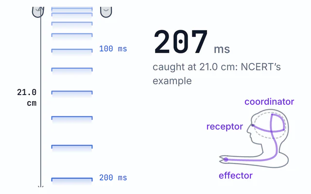
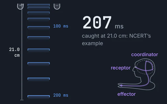
Tap the knee and watch the reflex arc in slow motion, from receptor to spinal cord to muscle, timed to the millisecond. Label the five parts or cut it.
Simulator
Tap the knee or drag the hammer back. Keys: T taps, N goes to the next event, arrows step the clock. Space plays and pauses.
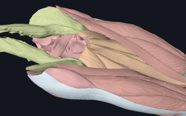
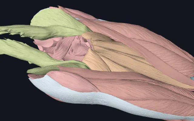
Leg drawn from BodyParts3D, © The Database Center for Life Science, CC BY 4.0. The nerves and cord are a diagram, not to scale.
A tap of 50 percent on the patellar tendon of a 175 cm adult. 8.7 ms: The Ia volley reaches the spinal cord through the dorsal root. 9.7 ms: The quadriceps’ alpha motor neurons fire, 30 of the pool of 100. 10.2 ms: The Ia inhibitory interneuron’s IPSP reaches the hamstrings’ motor neurons, so they stay quiet. 19.2 ms: The quadriceps EMG begins: the reflex latency. 29.2 ms: The quadriceps begin to pull, 10 ms after the EMG. 466 ms: The leg reaches its highest kick, 14.9 degrees of knee extension. Reflex present.
- Reflex latency From the hammer’s strike to the first deflection of the quadriceps EMG. 7.69 ms of it is the afferent volley, 8.33 ms the efferent, and the rest the receptor, one synapse, the motor neurons’ rise to threshold, the junction and the muscle fibres.
- 19.2 ms
- Peak knee extension Reached 466 ms after the tap. The shank swings as a physical pendulum, so the kick peaks long after the muscle’s force has.
- 14.9 °
- Motor neurons recruited Thresholds rise smallest cell first over a factor of ten (Henneman’s size principle). The volley’s drive here is 2.00 times the lowest threshold, with excitability 1.00.
- 30 of 100
- Central synapses One central synapse to the quadriceps: the knee jerk is monosynaptic. The hamstrings are inhibited through one interneuron, so the branch to the hamstrings has 2 and is disynaptic.
- 1 monosynaptic
- Afferent conduction
- 7.69 ms
- Efferent conduction
- 8.33 ms
- Peak force Reached 109 ms after the tap, from the muscle twitch simulator’s model, with an 80 ms contraction time and a 10 ms electromechanical delay.
- 6.0 % of max
- Status
- Reflex present
A teaching model, not a clinical test. It does not grade or localise a real person’s reflexes.
- Ia afferent, entering the cord
- Quadriceps motor neuron
- Hamstring motor neuron
- Quadriceps EMG
- Knee extension
Citing this tool
Last updated . Add the date you accessed it as well, which a citation of a page that can change asks for. If a specific result matters, cite the permalink from the tool’s share row instead of this page: it reproduces the exact parameters.
On the syllabus: MCAT, 3A: Structure and functions of the nervous and endocrine systems and ways these systems coordinate the organ systems.
The equation
Definition of reflex latency: conduction time plus synaptic and junctional delays
What are the five anatomical components of a somatic reflex arc?
A somatic reflex arc (such as the patellar knee-jerk reflex) executes an involuntary, rapid motor response via five sequential anatomical components: a sensory receptor detecting the stimulus, an afferent sensory neuron transmitting signals through the dorsal root, an integrating centre in spinal grey matter, an efferent motor neuron leaving through the ventral root, and a peripheral effector muscle.
A reflex is a fast, automatic response to a stimulus, and the reflex arc is the pathway that produces it. A textbook arc has five parts: a receptor that detects the stimulus, a sensory neuron that carries the signal into the spinal cord, a relay neuron inside the cord, a motor neuron that carries the command out again, and an effector, the muscle or gland that responds. The arc is the wiring; the reflex action is what the wiring does, such as the kick when a doctor taps your knee.
This simulator follows three arcs through one picture, a seated left leg drawn from real muscle and bone meshes beside a cross-section of the spinal cord. The knee jerk is the stretch reflex: tap the patellar tendon and the quadriceps contract. Withdrawal is the flexor reflex: a pin in the sole and the leg pulls away. Crossed extensor adds the other leg to the withdrawal, and it straightens to take the weight. Every stage is timed to the millisecond, and the auto time base plays the first 40 ms of a knee jerk 100 times slower than real time, so you can watch the sensory signal climb to the cord and the motor signal come back.
Worked example: the 19.2 ms knee jerk, term by term
The reflex latency is the time from the hammer’s strike to the first sign of the quadriceps
firing on an EMG, the electrical signal that comes before any force. It is a sum of delays,
t = t_r + Σ L/v + n δ_syn + τ + δ_m: the receptor’s own delay, the time to conduct
along every stretch of nerve, one synaptic delay per synapse, the time the motor neurons take to
reach threshold, and the muscle’s own delay. At the defaults, for a 175 cm adult:
- Receptor and tap transmission: 1.0 ms for the tap to stretch the muscle spindles and for them to fire.
- Sensory conduction: 0.38 m of femoral nerve and 0.12 m of dorsal root,
0.50 m ÷ 65 m/s = 7.7 ms, so the volley reaches the cord at 8.7 ms. - One synapse: 0.5 ms, which starts the EPSP in the quadriceps’ motor neurons at 9.2 ms.
- Rise to threshold: 0.5 ms, so the motor neurons fire at 9.7 ms.
- Motor conduction: 0.12 m of ventral root and 0.38 m of femoral nerve,
0.50 m ÷ 60 m/s = 8.3 ms, reaching the neuromuscular junctions at 18.0 ms. - Neuromuscular junction: 0.7 ms for the transmitter to fire the muscle fibres.
- Muscle fibres: 0.5 ms for their action potential to reach the EMG electrode.
Adding them up, 1.0 + 7.7 + 0.5 + 0.5 + 8.3 + 0.7 + 0.5 = 19.2 ms. Conduction is
16.0 ms of it, 83 percent: most of a reflex is the signal travelling about a metre of nerve, out
and back. The muscle’s force begins 10 ms after the EMG, at 29.2 ms, the electromechanical delay,
and peaks at 6.0 percent of its maximum at 109 ms. The leg is slower still. It swings like a
pendulum, so the kick reaches its highest, 14.9 degrees, at 466 ms, long after the muscle’s pull
has peaked.
Taller people have longer nerves, so they have longer latencies. Every path here scales with height, so each centimetre adds 0.0916 ms: a 150 cm adult’s knee jerk takes 16.9 ms and a 200 cm adult’s 21.5 ms. Frijns and colleagues (1997), measuring patellar tendon reflexes in 102 controls, found the latency rising with height with a correlation of 0.70, and measured latencies sit either side of this model’s: Khatun and colleagues, recording from the rectus femoris of 40 healthy volunteers, found the response beginning on average 17.7 ms after the hammer struck in those under 30 and 20.9 ms in those over 50. Halve both conduction velocities to 32.5 and 30 m/s, as a severe demyelinating neuropathy might, and the conduction time doubles while every fixed delay stays put, so the latency becomes 35.3 ms.
Why the knee jerk is monosynaptic
The Ia sensory fibres from the quadriceps’ muscle spindles synapse directly on the quadriceps’ own motor neurons. There is one central synapse between the receptor and the muscle, and no relay neuron at all on that path, which is why the knee jerk is called monosynaptic and why it is so fast: a synapse costs only about half a millisecond, but a chain of interneurons adds time at every link. Raise the synaptic delay slider from 0.5 to 2 ms and the knee jerk slows only from 19.2 to 20.7 ms, because it has only one synapse to slow.
Many school textbooks, India’s class 10 science textbook among them, draw “the” reflex arc with a relay neuron between the sensory and motor neurons, and they are right about the reflex they draw: it is the withdrawal arc, the hand pulling back from a flame or a pin. Switch this simulator to Withdrawal and the relay neurons appear in the cord. The knee jerk is the exception that has none, and the Label the five parts switch marks the only relay neuron it does have, on the branch to the hamstrings.
Reciprocal inhibition
A kick works better if the muscle that opposes it relaxes. So the same Ia volley that excites the quadriceps also excites a Ia inhibitory interneuron, which inhibits the motor neurons of the hamstrings, the knee’s flexors. That branch has two synapses. Its IPSP reaches the hamstring motor neuron at 10.2 ms, exactly 1 ms after the quadriceps’ EPSP begins at 9.2 ms: one synaptic delay plus the interneuron’s own 0.5 ms to reach threshold. The first plot shows both, the quadriceps motor neuron firing and the hamstring motor neuron pushed down below its resting potential of −70 mV, to −73.0 mV at the defaults.
Withdrawal and the crossed extensor
In the withdrawal reflex the receptors are nociceptors in the skin of the sole, and their thinly myelinated A-delta fibres conduct at about 20 m/s. The volley has 1.15 m to travel, up the tibial and sciatic nerves to the dorsal horn, which takes 57.5 ms, so it arrives at 59.5 ms. Two excitatory interneurons relay it to the flexor motor neurons, three synapses in all, and the model gives the interneurons 25 ms to integrate their input. That figure is a calibration, chosen so the EMG of the hamstrings begins at 96.4 ms, inside the 90 to 180 ms window in which flexion reflex studies such as Lee and Yoon (2017) score the late, nociceptive RIII response. Sandrini and colleagues (2005) review the evidence that the RIII component is carried by A-delta fibres and an earlier RII component by A-beta fibres. The motor neurons fire a burst of 8 volleys at 60 Hz rather than one, so the flexors’ twitches sum to 24.0 percent of their maximum force, and the knee is pulled up furthest, 34.0 degrees, at 262 ms.
At the same time an inhibitory interneuron silences the quadriceps, so the knee can bend. In the crossed extensor mode commissural interneurons carry the volley across the midline, through one more synapse and 5 ms more integration, to the other leg’s extensors, whose EMG begins at 101.0 ms. While one leg pulls away, the other straightens to take the weight. Switch on the slow C fibres to see the second, burning pain: at about 1 m/s they need 1.15 s to reach the cord, and arrive at 1152 ms, long after the foot has left the pin.
Cutting the arc
Turn on Cut mode and tap any of the eight ringed sites. A cut stops the volley where it is: cut the sensory nerve and the volley runs up to the cut and no further, so the cord, the muscle and the leg all stay still, while everything before the cut is unchanged. Cut the ventral root and the motor neurons still fire, but their volley never reaches the muscle. Block the neuromuscular junction, as the arrow poison curare does, and the motor volley arrives to no effect: no EMG and no kick.
The descending pathway is different. In this model the brain’s descending input inhibits the motor neurons, so cutting it, as an upper motor neuron lesion does, makes them more excitable rather than silencing them: the same tap recruits 60 of the 100 motor neurons instead of 30, and the kick reaches 30.1 degrees. Jendrassik reinforcement, hooking the fingers together and pulling them apart while the knee is tapped, makes the reflex brisker too; here it makes the motor neurons 1.5 times as excitable, which recruits 48 of them and kicks the leg 23.9 degrees. How reinforcement works is still debated, so that factor is a stated simplification, not a measurement.
What absent or brisk reflexes tell a clinician
Because every part of the arc is needed, a reflex that is missing points to a break somewhere in it. A lesion of the L3 or L4 nerve root, or of the femoral nerve, can weaken or abolish the knee jerk, while damage above the spinal segment, which removes descending inhibition, can make it brisker than normal. Clinicians compare the two sides, use reinforcement before calling a reflex absent, and read reflexes alongside strength and sensation. This simulator is a teaching model, not a clinical test: it does not grade or localise a real person’s reflexes.
Reading the plots
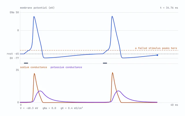
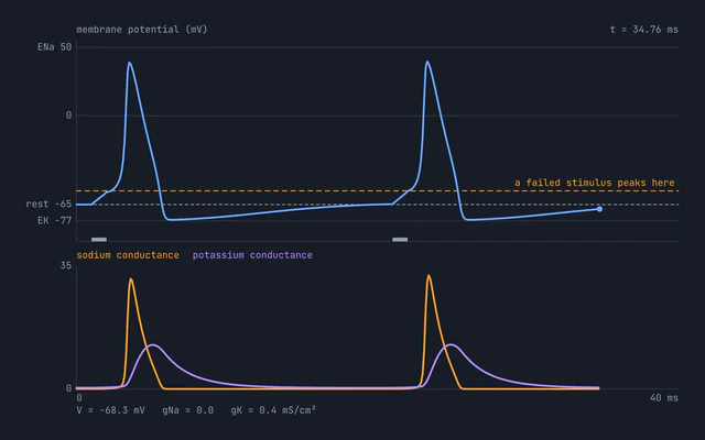
The first plot is the membrane potential of one representative cell of each population over the first 40 ms of a knee jerk: the Ia afferent where it enters the cord, the quadriceps motor neuron and the hamstring motor neuron. Each spike is the action potential the action potential simulator computes, Hodgkin and Huxley’s squid axon given 10 µA/cm² for 1 ms, moved so its rest reads −70 mV and compressed in time to 0.5 ms wide at half its amplitude, since the 1952 squid kinetics at 6.3 °C are several times slower than a warm mammalian axon. The resting level itself is the subject of the resting membrane potential simulator.
The second plot is the muscle’s EMG, a drawn compound potential sized by the share of motor neurons recruited, and the third is the knee’s angle. On their shared 800 ms axis the EMG is a flicker at 19 ms, over long before the leg has moved: the electrical events of a reflex are fast, and the mechanics are slow. Each plot downloads its numbers as CSV.
How the model works
The timetable is closed form: each arrival is the tap time plus the receptor’s delay, the length of each stretch of nerve over its conduction velocity and the delay at each synapse, with no approximation. The 100 motor neurons in each pool have thresholds spaced evenly on a logarithmic scale over a factor of ten, smallest cells first, the size principle Henneman described in 1957. The volley’s drive is proportional to the tap’s strength and to the pool’s excitability, and with descending inhibition intact and no reinforcement a tap below 25 percent brings none of them to threshold.
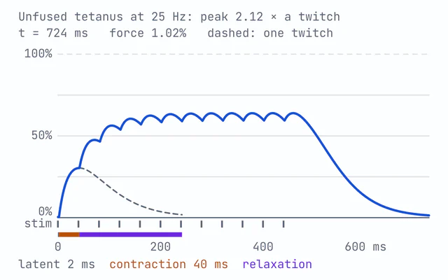
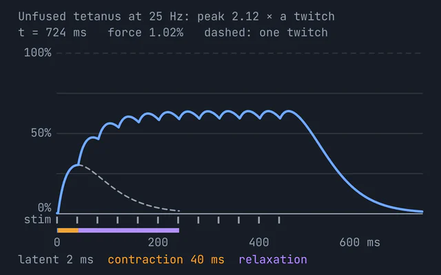
The muscle is the one in the muscle twitch and tetanus simulator, imported unchanged: a twitch with an 80 ms contraction time and a twitch to tetanus ratio of 0.2, scaled by the share of motor neurons recruited. The shank and foot swing as one physical pendulum about the knee, with the segment proportions Winter’s Biomechanics and Motor Control of Human Movement (2009) gives after Dempster and after Drillis and Contini: at 175 cm and 70 kg the segment is 0.4305 m long, weighs 4.27 kg and has a moment of inertia of 0.428 kg m² about the knee, so it swings with a natural period of 1.24 s. Its damping ratio of 0.2 and the quadriceps’ torque at full force, 64 N m, are calibrations that make a default tap kick about 15 degrees, not measurements: 64 N m is well under what a real quadriceps exerts at its strongest. The equation of motion is stepped by fourth-order Runge-Kutta at 0.5 ms, starting at the moment the quadriceps begin to pull, so that no step straddles the instant the force starts to rise.
The kick’s timing can be set against Mamizuka and colleagues (2007), who timed knee jerks with an accelerometer at the ankle. Their participants’ legs began to move 29.6 ms after the tap on average, and this one begins at 29.2 ms. The 10 ms electromechanical delay that puts it there is at the short end of measured values: Zhou and colleagues (1995) found about 22 ms between the EMG and the force of the knee extensors’ tendon reflex, measured with a load cell. The swing then gathers speed more slowly than a real one, taking 205 ms from the start of the movement to its fastest where Mamizuka’s participants took 150.8 ms, so a real kick may well peak sooner than this one’s 466 ms.
The leg is drawn from the left leg’s muscle and bone meshes in BodyParts3D, © The Database Center for Life Science, licensed under CC BY 4.0: the same meshes as the lower limb muscles explorer, where the rectus femoris drawn here can be turned in 3D. Each outline is the part of a mesh visible from the side, and the standing body is posed seated by turning the thigh about the centre of the femoral head and the shank about the knee. The nerves are not in the meshes, so they are drawn along the muscles as a diagram, and the spinal cord is drawn by hand.
What this model leaves out
- Spindle dynamics. A real spindle signals how fast it is stretched as well as how far, and falls silent while the muscle shortens. Here the spindle is a fixed 1.0 ms delay.
- Gamma motor neurons. They set a spindle’s sensitivity by tightening its own fibres, which is one way the brain sharpens or damps a reflex. They are not modelled.
- Mixed motor units. Every motor unit here shares one twitch, where a real muscle mixes fast and slow units, and every recruited cell takes the same 0.5 ms to reach threshold, where a stronger volley brings each one there a little sooner.
- Renshaw cells. Motor neurons inhibit themselves through Renshaw cells, which cut a burst short. There are none here.
- Cortical timing. A branch of the afferent volley climbs the cord to tell the brain, which is how you feel the tap. It is drawn as a dashed tract, but not timed.
- The withdrawing leg’s mechanics. In the withdrawal modes the legs follow the muscles’ force directly, with no inertia of their own.
Common mistakes
- Thinking the brain decides the kick. The motor neurons fire 1 ms after the volley reaches the cord, at 9.7 ms. The decision is made in the spinal segment, and the brain is told afterwards.
- Calling every reflex polysynaptic. The withdrawal reflex is, with three synapses to the flexors, but the knee jerk has one.
- Confusing the latency with the kick. The latency is measured to the EMG, 19.2 ms; here the leg does not reach its highest point until 466 ms.
- Thinking a stronger tap is a faster reflex. Here a harder tap recruits more motor neurons and kicks higher, but every delay on the path is the same, so the latency does not change.
- Reading an inhibited muscle as a contracting one. Reciprocal inhibition keeps the hamstrings quiet; it does not pull the knee the other way.
Model and assumptions
- Method
- Runge-Kutta 4th order
- Fixed step
- 0.5 ms
- Repeatability
- Deterministic. The same link gives the same numbers on any machine.
What it assumes
- Every arrival is a closed-form sum: the receptor’s delay, each stretch of nerve’s length over its conduction velocity, one synaptic delay per synapse and a fixed time for the cells that must sum their inputs to threshold.
- Every path is written for a 175 cm adult and scaled by height over 175, so a taller person’s nerves are longer in proportion: the conduction times change and no other delay does.
- Each population is one representative cell, and every spike is the Hodgkin and Huxley waveform the action potential simulator computes, moved so rest reads −70 mV and compressed to 0.5 ms wide at half its amplitude.
- The 100 motor neurons in a pool have thresholds spaced evenly on a logarithmic scale over a factor of ten, so a harder tap or a more excitable pool recruits more of them, smallest first.
- The muscle’s force is the muscle twitch simulator’s twitch, with an 80 ms contraction time and a 10 ms delay from EMG to force, scaled by the share of the pool recruited.
- The shank and foot swing as one rigid pendulum about the knee with Winter’s segment proportions, stepped by RK4 at 0.5 ms from the moment the quadriceps begin to pull; its damping ratio of 0.2 and the 64 N m torque at full force are calibrations.
- In the withdrawal modes the legs follow the muscles’ force along a smooth curve towards a fixed limit, with no dynamics of their own, and the 25 ms the interneurons take to integrate is a calibration.
Where it stops holding. Where a reflex depends on more than its delays. Spindle dynamics, gamma motor neurons, Renshaw cells and mixed motor units are not modelled, so the size and shape of a real response, and how it changes with how fast the tendon is stretched, can differ from these. Where the shape of a single spike matters, its currents and its refractory period, this model only borrows a fixed waveform. The Action Potential Simulator is the right tool there.
Numerical accuracy
- Estimated error
- 1.4e-12 rad in the knee angle, about 5.2e-12 of the largest value reached
- How that was obtained
- Running the same problem again at half the step changed the answer by at most 1.3e-12 rad over 3.17 s, from the quadriceps’ first pull to the end of the run. Richardson extrapolation of that difference gives the figure above.
- Observed order
- 3.99, measured from a second halving rather than assumed
- Conditions
- a 50 percent tap on the patellar tendon of a 175 cm, 70 kg adult, descending inhibition intact, the shipped defaults
Common questions
What are the five parts of a reflex arc?
A receptor that detects the stimulus, a sensory neuron that carries the signal into the spinal cord, a relay neuron inside the cord, a motor neuron that carries the command back out, and an effector, the muscle or gland that responds. That is the withdrawal arc, the one most textbook diagrams draw, with the hand pulling back from a flame or a pin. The knee jerk is the exception: its sensory neurons synapse directly on its motor neurons, so its main path has no relay neuron at all.
Why is the knee-jerk reflex called monosynaptic?
Because there is only one central synapse between the receptor and the muscle. The Ia sensory fibres from the quadriceps’ muscle spindles synapse directly on the quadriceps’ own motor neurons, so the signal crosses a single junction in the spinal cord. The same fibres also excite a Ia inhibitory interneuron, which inhibits the motor neurons of the hamstrings so they do not resist the kick. That branch has two synapses, and in this model its inhibition reaches the hamstrings’ motor neurons 1 ms after the quadriceps’ excitation begins: one synaptic delay of 0.5 ms and 0.5 ms for the interneuron to reach threshold.
How fast is the knee-jerk reflex?
About 20 ms from the tap to the first sign of the quadriceps firing on an EMG. Khatun and colleagues, recording from the rectus femoris of 40 healthy volunteers, found the response beginning on average 17.7 ms after the hammer struck in those under 30 and 20.9 ms in those over 50. In this model a 175 cm adult’s takes 19.2 ms, and 16.0 ms of that, 83 percent, is the signal conducting along about a metre of nerve, to the spinal cord and back. Taller people have longer nerves and longer latencies, as Frijns and colleagues found in 1997, and here each centimetre of height adds 0.0916 ms. The leg itself is far slower: here the kick is highest at about 466 ms.
Does the brain take part in a reflex?
Not in the arc itself. The motor neurons in the spinal cord fire 1 ms after the sensory signal arrives there, and nothing has to reach the brain and come back for the kick to happen. A branch of the same signal does climb the cord to the brain, which is how you feel the tap, but it gets there after the decision has been made. The brain’s part is to set how brisk the reflex is: descending pathways hold the motor neurons back, and in this model removing that inhibition, as an upper motor neuron lesion does, doubles the share of motor neurons the same tap recruits, from 30 of 100 to 60.
What is the crossed extensor reflex?
The other half of a withdrawal. When one foot steps on something sharp, its flexor muscles pull it away, and at the same time commissural interneurons carry the signal across the spinal cord to the other leg, exciting its extensors and inhibiting its flexors, so that leg straightens and takes the weight. In this model the pricked leg’s hamstrings show EMG at 96.4 ms and the other leg’s quadriceps at 101.0 ms, after one more synapse and 5 ms more integration in the cord.
What happens if the reflex arc is cut?
The reflex is lost. Every part from the receptor to the neuromuscular junction is needed, so a break anywhere stops the signal there and nothing beyond it happens, which is why damage to the L3 or L4 nerve root or to the femoral nerve can weaken or abolish the knee jerk. A blocked junction, as the arrow poison curare causes, is the odd one out: the motor volley still arrives, but the muscle never responds. Damage above the spinal segment does the opposite, because it removes descending inhibition and makes the reflex brisker. This simulator is a teaching model, not a clinical test.
What is the difference between a reflex arc and a reflex action?
The reflex arc is the pathway: the receptor, sensory neuron, relay neuron, motor neuron and effector, wired in a chain. The reflex action is what that pathway does, the fast, automatic response it produces, such as the kick of the leg when the patellar tendon is tapped or the hand jerking back from a hot pan.