Dermatome and Myotome Map Explorer
Click a spinal root from C2 to S5 to light its dermatome on a front and back body map, with its myotome muscles, key sensory point and tendon reflex.
Visualiser
With the map focused, the up and down arrow keys step from root to root, and Home and End go to C2 and S5.
Whole body view. C6, the outer forearm, the thumb and the index finger, is lit.
Cervical spinal root
C6:The outer forearm, the thumb and the index finger
- Key sensory point
- The back of the thumb’s proximal phalanx
- Key movement
- Wrist extension
- Reflex
- Biceps reflex, brachioradialis reflex and triceps reflex, in part
- Usual disc
- C5/C6
Lesion
Reflex
Muscles C6 supplies
See them in 3DMajor supply Minor supply
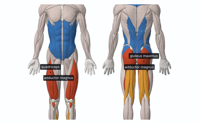
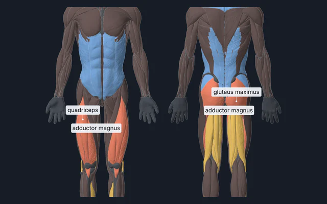
Major serratus anterior, subscapularis, teres major, pectoralis major, clavicular part, biceps brachii, brachialis, coracobrachialis, brachioradialis, supinator, pronator teres, extensor carpi radialis longus, extensor carpi radialis brevis and flexor carpi radialis.
Minor deltoid, supraspinatus, infraspinatus, teres minor, pectoralis major, sternal part, latissimus dorsi and triceps brachii.
Outlines drawn from BodyParts3D, © The Database Center for Life Science, CC BY 4.0.
A teaching map. Root territories overlap and vary between people, and a root lesion is diagnosed by examination and imaging, not by a map. Numbness between the legs or around the anus, new bladder or bowel problems, or quickly spreading weakness need emergency care.
Citing this tool
Last updated . Add the date you accessed it as well, which a citation of a page that can change asks for. If a specific result matters, cite the permalink from the tool’s share row instead of this page: it reproduces the exact parameters.
What is a dermatome and myotome map?
A dermatome is a specific area of skin sensory innervation supplied by a single spinal nerve root (from C2 to S5), while a myotome is the group of skeletal muscles innervated by motor fibres of a specific spinal root. Dermatome maps are vital for localising spinal cord injuries and radiculopathies.
Dermatomes and myotomes in one picture
A root’s band of skin and its muscles come from the same segment of the embryo, so they are examined together: a root that is pressed on numbs its own band of skin, weakens the movements it mainly drives and dulls the reflex it mainly carries.
The map draws the 29 spinal roots that have a dermatome, C2 to C8, T1 to T12, L1 to L5 and S1 to S5. C1 has none in most people, because it usually has no sensory root, and S4 and S5 are drawn as one ring round the anus, since the standard examination tests them together. The face is not a spinal territory at all: it is the trigeminal nerve’s, and the map labels its three divisions as a cranial nerve’s, V1, V2 and V3. Choose a root and the muscles it supplies light on the flat muscle map under it, the major supply solid and the minor supply tinted, and the reflex it carries is tapped.
The key points every examination uses
A border between two dermatomes is never sharp, so examinations test each root at one point well inside its band. The International Standards for Neurological Classification of Spinal Cord Injury, the ISNCSCI of the American Spinal Injury Association, fix one key sensory point a side for each of the 28 levels from C2 to S4 and S5, tested with light touch and with a pin, and one key movement for each of C5 to T1 and L2 to S1. The chosen root’s key point is always ringed, with the root’s name pinned beside it; turn on Every key sensory point and the rest appear as smaller rings where the standard puts them. The table at the end lists every one, read from the same data as the map.
A few are worth knowing by heart: the back of the thumb’s proximal phalanx for C6, the nipple line for T4, the umbilicus for T10, the medial malleolus, the inner ankle bone for L4 and the outer side of the heel for S1.
Worked example: a C5/C6 disc and the C6 root
Choose C5/C6 under Lesion. The map moves to C6 and hatches its band, and the Lesion panel says what that does, and why:
- Sensation. Numbness or tingling in the C6 band: the outer forearm, the thumb and the index finger. Usually a smaller patch than the whole band, because C5 and C7 overlap it: Sherrington found in 1893 that cutting a single root in monkeys left no skin entirely without feeling.
- Strength. Wrist extension, the standard’s key movement for C6 (extensor carpi radialis longus and extensor carpi radialis brevis). Also weaker where C6 is a major supply: serratus anterior, subscapularis, teres major, pectoralis major, clavicular part, biceps brachii, brachialis, coracobrachialis, brachioradialis, supinator, pronator teres and flexor carpi radialis.
- Reflexes. The biceps reflex reduced or absent, the brachioradialis reflex reduced or absent and the triceps reflex may be reduced.
- Why C6 and not C5. Cervical roots leave the spine above the vertebra of the same number, so the C6 root leaves between the C5 and C6 vertebrae, right beside the C5/C6 disc. A disc bulging backwards and to the side presses on it there, which is why a C5/C6 disc gives C6 symptoms.
In the lumbar spine the same disc gives a different root. A typical L4/L5 herniation presses on L5, and one far out to the side catches L4 instead; the Far-lateral herniation option shows both.
Why maps disagree and overlap
Dermatome maps in textbooks differ, sometimes by a whole level, because they were made in different ways. Foerster (1933) mapped the sensation left over in patients after several roots above and below one root had been cut. Keegan and Garrett (1948) mapped the numbness of patients whose single roots were compressed by discs, and drew long continuous bands down the limbs. Lee, McPhee and Stringer (2008) reviewed the evidence behind the maps in use, found much of it flawed, and drew only the areas that were consistent in most people, leaving the skin between them blank.
The map here is our own drawing, not a copy of any of these. Its bands run on until they meet, so that any patch of skin can be tapped, and each holds its own key sensory point from the standard; where two bands meet, the border is a line on a drawing, not one found on a body. It also parts from Lee’s map in places: the inner forearm, most of which this map gives to C8, is T1 in theirs, as it is in Foerster’s.
Neighbouring dermatomes also overlap. Sherrington found in 1893 that cutting a single root in monkeys left no skin entirely without feeling, only small patches where it was dulled, because the roots above and below it still reached its band. That is why one root’s lesion usually numbs a smaller patch than its band, and why the map feathers the chosen band’s edge into its neighbours. Turn on Where maps disagree to see the areas the published maps give to different roots, such as the index finger, the big toe and the sole.
Reflexes and their roots
A tendon reflex is a stretch reflex: the tap stretches the muscle, its stretch receptors fire, and the signal enters the cord through one to three dorsal roots and returns to the same muscle through the matching ventral roots. A root lesion breaks that loop, so the reflex it mainly carries is reduced or absent, while a lesion of the cord above it does the opposite and makes reflexes brisk. The six limb reflexes the map taps are below, with every root that Walker (1990) or StatPearls lists for each and, as the main one, the root the MSD Manual names. The face has one more, the jaw jerk, a stretch reflex of the chewing muscles that runs through V3; the map does not draw it.
| Reflex | Roots | Mainly | Tap | Response |
|---|---|---|---|---|
| Biceps reflex | C5, C6 | all of them | the biceps tendon in the elbow crease, through the examiner’s thumb | the elbow bends |
| Brachioradialis reflex | C5, C6 | C6 | the thumb side of the forearm a few centimetres above the wrist | the elbow bends and the forearm turns palm up |
| Triceps reflex | C6, C7, C8 | C7 | the triceps tendon just above the point of the elbow | the elbow straightens |
| Finger flexor reflex | C8, T1 | C8 | the examiner’s fingers laid across the patient’s slightly bent fingers | the fingers curl |
| Knee jerk | L2, L3, L4 | L4 | the patellar tendon below the kneecap, with the leg hanging | the knee straightens with a kick |
| Ankle jerk | S1, S2 | S1 | the Achilles tendon, with the foot held slightly up | the foot points down |
Dermatomes of the upper limb
The roots run round the arm in order, from the thumb’s side to the little finger’s. C5 is the outer upper arm, over the deltoid, down to the elbow; C6 is the outer forearm, the thumb and the index finger; C7 is the middle finger, and a strip down the back of the forearm; C8 is the ring and little fingers, the inner edge of the hand and the inner forearm; and T1 is the inner side of the arm and elbow, and the top of the inner forearm, with T2 above it in the armpit. Because C5 to T1 are carried out into the limb, the chest has C4 directly above T2, the ventral axial line the Upper limb view shows. The key points are the outer and inner sides of the elbow crease for C5 and T1, and the backs of the thumb, middle and little fingers for C6, C7 and C8.
Dermatomes of the hand
The Hand view draws the palm and the back of the right hand. The thumb and index finger are C6, the middle finger C7, and the ring and little fingers C8, on both sides. On the palm, C7 reaches only the middle of the palm, so at the wrist C6 meets C8; on the back of the hand it runs down to the wrist. The index and ring fingers are where maps differ most.
Dermatomes of the lower limb
The lower limb turned inwards as it grew, so its bands spiral. L1 is the groin, just below the inguinal ligament, and the upper buttock; L2 is the front and outer thigh, running down and in; L3 is the lower front of the thigh and the inner knee; L4 is the inner shin, the inner ankle and the inner edge of the foot; L5 is the outer shin, the top of the foot and the big toe; S1 is the outer edge and sole of the foot, the heel and the lower back of the calf; and S2 is a strip down the back of the thigh and calf, through the back of the knee. S3 to S5 are rings round the anus, the saddle area. The key points are the inner ankle for L4, the top of the foot at the third toe joint for L5, the outer heel for S1 and the back of the knee for S2.
Dermatomes of the foot
The Foot view draws the top of the right foot and its sole. On top, L4 runs along the inner border to the base of the big toe, L5 covers the rest of the top with the first four toes, and S1 the outer border and the little toe. Underneath, S1 has the heel and the outer sole, L5 the inner forefoot, and L4 a strip along the arch; the inner sole is the part of the foot the maps agree on least.
Dermatomes of the face and neck
The face is not divided into spinal dermatomes. The trigeminal nerve supplies it in three divisions: V1, the ophthalmic, the forehead and scalp to the crown, the upper eyelid and the bridge and tip of the nose; V2, the maxillary, the lower eyelid, the cheek, the side of the nose and the upper lip; and V3, the mandibular, the lower lip, the chin, the jaw except its angle, the temple and the front of the ear. The angle of the jaw is not trigeminal: it belongs to C2 and C3 through the great auricular nerve. C2 is the back of the scalp, behind the ear and the angle of the jaw, C3 the neck, front and back, down to the collarbones, and C4 the tops of the shoulders and a cape across the upper chest and back. The Face and neck view draws the face from the front and the head from the side.
What this map leaves out
- Peripheral nerve territories. The median, ulnar and radial nerves each carry fibres from several roots, so their areas of skin differ from dermatomes: numbness of the thumb, index and middle fingers from carpal tunnel syndrome is a median nerve pattern, not a C6 or C7 one. Telling the two apart is a large part of examining a numb hand.
- Variation between people. Bands shift by part of a level from person to person, and a prefixed or postfixed brachial plexus moves the whole arm’s supply up or down by one root.
- Upper motor neuron signs. The lesion mode is a root, a lower motor neuron lesion. Damage to the spinal cord itself gives brisk reflexes, increased tone and an upgoing plantar response below the level, with sensory loss below a level rather than in one band.
- The coccygeal root and C1. Neither is drawn: C1 usually has no dermatome and the coccygeal area is too small to draw on a whole body.
Every root at a glance
All 29 spinal roots, with the dermatome, the standard’s key sensory point and key movement, the reflex each carries and the disc that usually presses on it, from the same data the map is drawn from.
| Root | Dermatome | Key sensory point | Key movement | Reflex | Usual disc |
|---|---|---|---|---|---|
| C2 | The back of the scalp, behind the ear and the angle of the jaw | At least 1 cm to the side of the occipital protuberance, or 3 cm behind the ear | None graded | None | None: no disc lies between the first and second vertebrae |
| C3 | The neck, front and back, down to the collarbones | The supraclavicular fossa, behind the collarbone at the midclavicular line | None graded | None | C2/C3 |
| C4 | The tops of the shoulders and a cape across the upper chest and back | The top of the acromioclavicular joint | None graded | None | C3/C4 |
| C5 | The outer upper arm, over the deltoid, down to the elbow | The outer side of the elbow crease, just above it | Elbow flexion | Biceps reflex and brachioradialis reflex, in part | C4/C5 |
| C6 | The outer forearm, the thumb and the index finger | The back of the thumb’s proximal phalanx | Wrist extension | Biceps reflex, brachioradialis reflex and triceps reflex, in part | C5/C6 |
| C7 | The middle finger, and a strip down the back of the forearm | The back of the middle finger’s proximal phalanx | Elbow extension | Triceps reflex | C6/C7 |
| C8 | The ring and little fingers, the inner edge of the hand and the inner forearm | The back of the little finger’s proximal phalanx | Finger flexion, at the tip of the middle finger | Finger flexor reflex and triceps reflex, in part | C7/T1 |
| T1 | The inner side of the arm and elbow, and the top of the inner forearm | The inner side of the elbow crease, just above the medial epicondyle | Little finger abduction | Finger flexor reflex, in part | T1/T2, rarely |
| T2 | The armpit, the inner upper arm and a band across the upper chest and back | The top of the armpit | None graded | None | T2/T3, rarely |
| T3 | A band round the chest at the third rib | The third intercostal space, at the midclavicular line | None graded | None | T3/T4, rarely |
| T4 | A band round the chest at the nipples | The fourth intercostal space, the nipple line | None graded | None | T4/T5, rarely |
| T5 | A band round the chest just below the nipples | The fifth intercostal space, midway between T4 and T6 | None graded | None | T5/T6, rarely |
| T6 | A band round the trunk at the lower end of the breastbone | The sixth intercostal space, level with the xiphisternum | None graded | None | T6/T7, rarely |
| T7 | A band round the trunk along the lower ribs | The seventh intercostal space, midway between T6 and T8 | None graded | None | T7/T8, rarely |
| T8 | A band round the trunk halfway from the breastbone to the umbilicus | The eighth intercostal space, midway between T6 and T10 | None graded | None | T8/T9, rarely |
| T9 | A band round the trunk just above the umbilicus | The ninth intercostal space, midway between T8 and T10 | None graded | None | T9/T10, rarely |
| T10 | A band round the trunk at the umbilicus | The tenth intercostal space, level with the umbilicus | None graded | None | T10/T11, rarely |
| T11 | A band round the trunk below the umbilicus | The eleventh intercostal space, midway between T10 and T12 | None graded | None | T11/T12, rarely |
| T12 | The lower abdomen above the groin crease, and the flank above the hip | The midpoint of the inguinal ligament | None graded | None | T12/L1 |
| L1 | The groin, just below the inguinal ligament, and the upper buttock | Halfway between the T12 and L2 points | None graded | None | T12/L1, or a far-lateral L1/L2 |
| L2 | The front and outer thigh, running down and in | The front inner thigh, halfway from the midpoint of the inguinal ligament to the medial femoral condyle | Hip flexion | Knee jerk, in part | L1/L2 |
| L3 | The lower front of the thigh and the inner knee | The medial femoral condyle, just above the knee | Knee extension | Knee jerk, in part | L2/L3 |
| L4 | The inner shin, the inner ankle and the inner edge of the foot | The medial malleolus, the inner ankle bone | Ankle dorsiflexion | Knee jerk | L3/L4, or a far-lateral L4/L5 |
| L5 | The outer shin, the top of the foot and the big toe | The top of the foot at the third metatarsophalangeal joint | Big toe extension | None | L4/L5, or a far-lateral L5/S1 |
| S1 | The outer edge and sole of the foot, the heel and the lower back of the calf | The outer side of the heel | Ankle plantarflexion | Ankle jerk | L5/S1 |
| S2 | A strip down the back of the thigh and calf, through the back of the knee | The middle of the popliteal fossa, behind the knee | None graded | Ankle jerk, in part | None on its own: inside the sacrum, reached only by a large central disc higher up |
| S3 | A ring round the anus on the inner buttocks, at the fold under the buttock | The ischial tuberosity, at the fold under the buttock | None graded | None | None on its own: inside the sacrum, reached only by a large central disc higher up |
| S4 | The skin round the anus, with S5 | Beside the anus, within 1 cm of its margin, tested with S5 | None graded | None | None on its own: inside the sacrum, reached only by a large central disc higher up |
| S5 | The skin round the anus, with S4 | Beside the anus, within 1 cm of its margin, tested with S4 | None graded | None | None on its own: inside the sacrum, reached only by a large central disc higher up |
Where the roots leave the spine
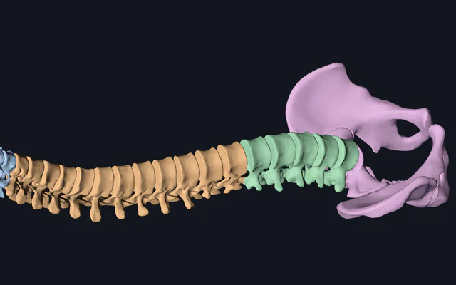
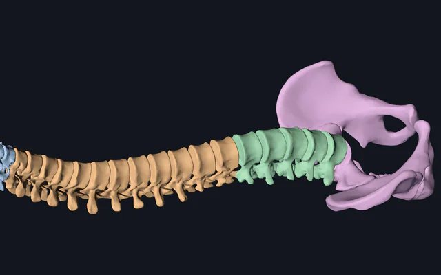
The roots leave through the openings between the vertebrae, which the vertebral column explorer shows in 3D, with the neck’s in the cervical spine and skull base explorer and the lower back’s in the lumbar spine and pelvis explorer. The skin itself is a single surface in the body surface explorer, and what one sensory fibre carries is an action potential, which the action potential simulator builds. The muscles each root supplies are in the upper limb and lower limb muscle explorers, and the muscles worked explorer colours a whole body by root in its root mode.
Related clinical and neuroanatomy simulators
Explore the neurological pathways, spinal anatomy and muscle actions that connect with each spinal root:
- Reflex Arc Simulator: trace sensory, interneuron and motor impulses through the spinal cord.
- Vertebral Column in 3D: all 24 articulating vertebrae where nerve roots exit intervertebral foramina.
- Action Potential Simulator: ion currents and impulse propagation in sensory and motor neurons.
- Muscles Worked by Exercise: whole-body 3D muscle maps and nerve root innervations.
- Dermatomes by nerve root: a page for each spinal root from C2 to S5, with its key sensory point, myotome, reflexes and usual disc, and for V1 to V3 of the face.
Common questions
What is a dermatome, and what is a myotome?
A dermatome is the area of skin whose sensation reaches the spinal cord through one spinal nerve root, and a myotome is the group of muscles one root supplies. Each of the 29 roots from C2 to S5 has a dermatome, while C1 usually has none, and the map lights a root’s band of skin, its muscles and its reflex together.
What are the dermatomes of the upper limb?
Running round the arm from the thumb’s side, C5 covers the outer upper arm, C6 the outer forearm, thumb and index finger, C7 the middle finger, C8 the ring and little fingers and the inner forearm, and T1 the inner arm and elbow. The standard test points are the outer side of the elbow crease for C5, the backs of the thumb, middle and little fingers for C6, C7 and C8, and the inner side of the elbow crease for T1.
What are the dermatomes of the lower limb?
L1 is the groin, L2 the front of the thigh, L3 the lower thigh and inner knee, L4 the inner shin and inner ankle, L5 the outer shin and the top of the foot, S1 the outer foot, sole and heel, and S2 a strip down the back of the thigh. The test points are the inner ankle for L4, the top of the foot at the third toe joint for L5 and the outer heel for S1.
Are there dermatomes on the face?
Not spinal ones. The face is supplied by the three divisions of the trigeminal nerve: V1 the forehead, upper eyelid and nose, V2 the cheek, lower eyelid and upper lip, and V3 the lower lip, chin and jaw, though not the angle of the jaw, which belongs to C2 and C3. The back of the scalp is C2.
Which reflex tests which nerve root?
The biceps reflex tests C5 and C6, the brachioradialis mainly C6, the triceps mainly C7, the finger flexors mainly C8, the knee jerk mainly L4 with L2 and L3, and the ankle jerk mainly S1. A root lesion reduces or abolishes the reflex it mainly carries, which the lesion mode shows.
Why do dermatome maps look different in different books?
They were made in different ways. Foerster (1933) mapped the sensation left after cutting neighbouring roots, Keegan and Garrett (1948) mapped numbness from disc herniations, and Lee, McPhee and Stringer (2008) reviewed the evidence and drew only the areas consistent in most people, leaving gaps between them. The map here is our own drawing, with no gaps, and in places it differs from theirs. Neighbouring dermatomes also overlap, which is why a single root lesion usually numbs a smaller area than its band.
Which nerve root does a C5/C6 or L4/L5 disc herniation affect?
Usually C6 and L5. Cervical roots leave the spine above the vertebra of the same number, so the C5/C6 disc meets the C6 root, while a typical lumbar herniation catches the root passing down to the next level, so L4/L5 compresses L5; one far out to the side catches L4 instead. Numbness between the legs, new bladder or bowel trouble, or rapidly worsening weakness needs emergency care.