Muscle Origin, Insertion and Action Explorer
Pick a muscle to see its origin and insertion painted on real 3D bones, with its action and nerve, or pick a bone to see the muscles attached to it.
Visualiser
- Origin
- Insertion
- Line of pull
Seen from the outer side
Shoulder and arm bones, 0.6 MB. Turn them, zoom in and click any bone.
Start here
- Find a muscle Type cuff, biceps or jaw to pick from 29 muscles of the shoulder, arm, jaw and neck. Its bones are drawn from the side that shows it best, origins red and insertions blue, numbered as the card lists them with the area each covers, its main action as a moving figure and its nerve.
- Open in 3D The real bones load in the same box with the same attachments painted and arrows from each insertion to its origins. Point at a row in the card, or a patch in the drawing, to light it in both.
- By bone Pick the scapula, the humerus or another bone and every muscle that arises from it or inserts on it is listed, origins first, and painted where it was measured. In 3D, click any bone.
Bone from five BodyParts3D skeletal models, and muscle from three when you ask for it. No ligaments, fasciae, aponeuroses or joint capsules, so an attachment to one, such as the trapezius’s to the ligamentum nuchae, is named in the card but not drawn, and where a muscle stops short of its bone, as at several vertebral spines, the site is a hollow pin. No muscles of the wrist, hand, hip or leg. Each patch is where this one body’s meshes meet its bones, not a textbook outline.
Where this comes from
Segmented from MRI of one adult male volunteer, then refined by medical illustrators, who filled in missing detail and sharpened blurred outlines from textbooks and atlases. Two consequences worth knowing: it is one person’s anatomy rather than an average, and faint stepping on some surfaces is the resolution of the original scan rather than a rendering fault.
Meshes from BodyParts3D, © The Database Center for Life Science, under Creative Commons Attribution 4.0 International. Names and identifiers are the dataset’s own: Foundational Model of Anatomy identifiers and English labels, as its parts list gives them. Converted meshes are shared under the same licence.
Changes made: Meshes were converted from binary STL to an indexed mesh with 16-bit quantised vertex positions, and surface normals were recomputed from the geometry. No vertex was moved other than by that quantisation, whose worst-case error on any one coordinate is reported on the page. On this page it is at most 0.00558 mm, the largest among the 8 models the page can load. The attachment footprints are ScienceQuest’s measurement of these meshes: the bone triangles each muscle’s meshes come within 1, 2, 3 mm of, inside a curated zone on the bone. The drawings the explorer opens on are the same meshes and footprints, projected from a fixed side and shaded by their own surface normals. Each model’s file is then compressed without loss, by meshoptimizer’s vertex and index codecs and gzip, and restored exactly before it is drawn.
Latin terms for 183 of the 399 structures in these models come from English Wikipedia anatomy infoboxes, under Creative Commons Attribution-ShareAlike 4.0, joined by Wikidata property P1402, Foundational Model of Anatomy identifier. Only a term a source states for that structure is used, never one constructed from a family name, which is why the rest carry no Latin rather than a guess.
Citing this tool
Last updated . Add the date you accessed it as well, which a citation of a page that can change asks for. If a specific view matters, cite the page address as it stands in your browser instead: it records what is selected and shown.
What do muscle origin, insertion, and action mean?
In musculoskeletal anatomy, a muscle’s origin is the attachment that remains relatively fixed during contraction (typically proximal or medial), while its insertion is the movable attachment drawn towards the origin. A muscle’s action is the movement that pull produces at the joints it crosses, such as flexion, extension, abduction or rotation.
A muscle pulls on the bones at both its ends. Its origin is the attachment that usually stays fixed, often the end nearer the trunk, and its insertion the one that moves; contraction draws the insertion towards the origin, which is why every arrow on the model points from an insertion back to its origin. The roles are a convention about which end moves, not a property of the muscle: in a pull-up the hand and the humerus are fixed and latissimus dorsi pulls the trunk up to the arm, insertion fixed and origin moving.
The explorer has 29 muscles of the shoulder and arm, the jaw and the neck, with 110 attachment sites between them, described as Gray’s Anatomy (1918) describes them, in modern terms. 96 are painted on the bone, 6 are pinned because the muscle and the bone do not quite meet in the scan, and 8 are on structures the dataset does not segment, such as the ligamentum nuchae and the thoracolumbar fascia, which are named rather than drawn.
How the patches were measured
Every structure here is part of one model of one man, segmented from MRI and refined by medical illustrators, in one coordinate frame, so where a muscle meets a bone can be measured from the meshes rather than drawn. For each site, every vertex of the bone is measured against the muscle’s meshes, exactly, and a bone triangle is painted when all three of its corners lie within a threshold and its centre lies in a zone curated for that site. The threshold starts at 1 mm and steps up to 3 mm when a footprint comes to less than 25 mm², because the ends of tendons often stop short of bone in the scan; still under that, a site is shown as a pin.
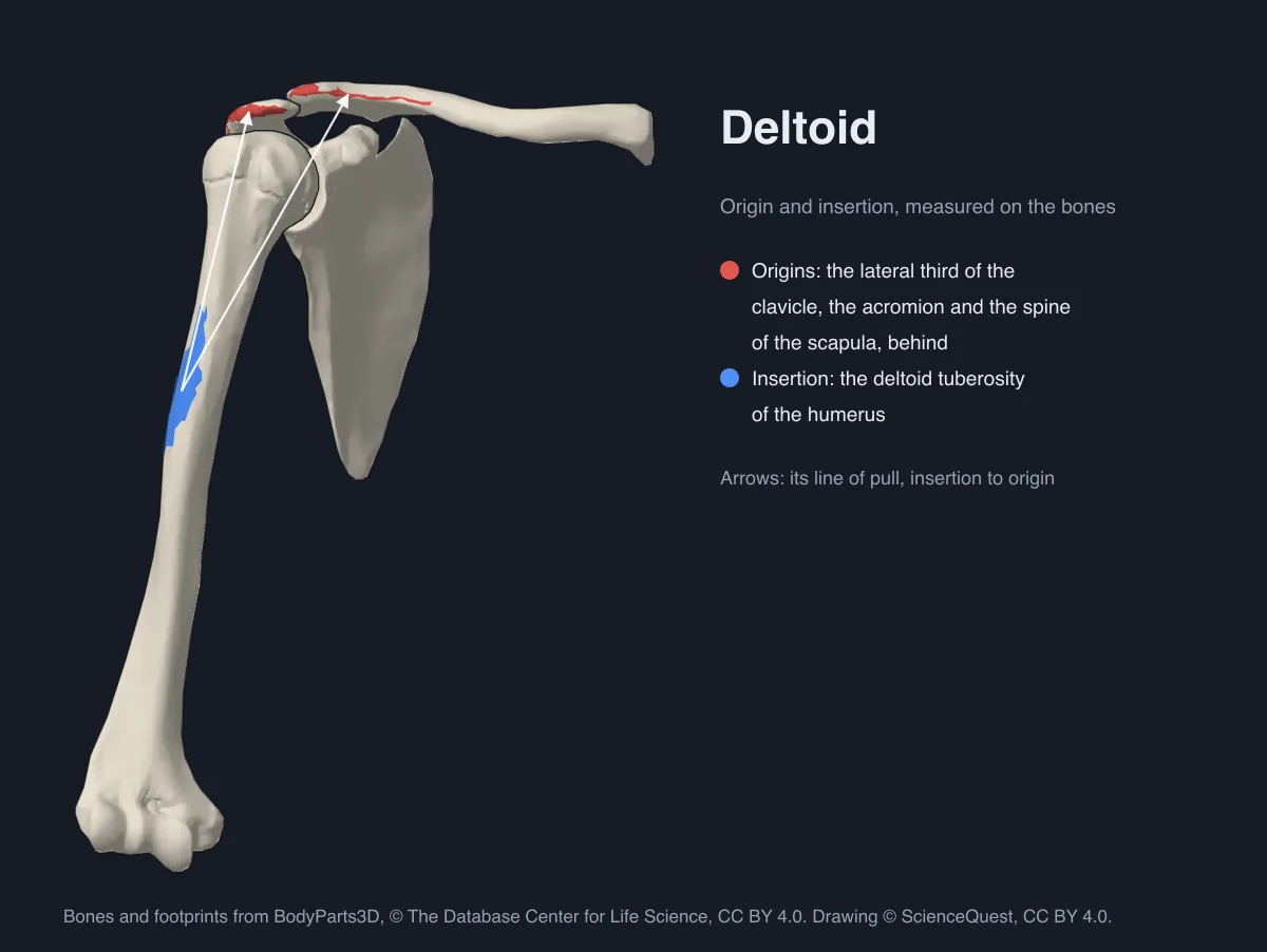
The zone is there because contact is not attachment. Within 1 mm the deltoid touches the humerus over 10.8 cm², but only 7.1 cm² of that lies on the outer side of the shaft, at the deltoid tuberosity, centred 37 percent of the way down; the rest is where the muscle wraps over the greater tubercle on its way there. The card gives each site’s area, and pointing at the area shows the threshold it took.
The rotator cuff
Four muscles from the scapula hold the head of the humerus in its shallow socket. Supraspinatus, infraspinatus and teres minor insert in that order down the three facets of the greater tubercle, centred 3, 6 and 11 percent of the way down the humerus in this body, and subscapularis, from the front of the scapula, on the lesser tubercle, centred 7 percent down. Type cuff in the picker to list all four.
Which muscles attach to the humerus
Choose By bone and pick the humerus, or click it in the model: of the muscles here, 6 arise from it and 9 insert on it, each painted on its front and back and listed in the card. The shoulder muscles insert on its upper end and shaft, brachialis and two heads of triceps arise from its shaft, and brachioradialis and the forearm’s rotators from its lower end and epicondyles.
The muscles of mastication
Masseter runs from the zygomatic arch to the outer surface of the ramus and angle of the mandible, temporalis from the temporal fossa to the coronoid process, medial pterygoid from the lateral pterygoid plate, the palatine bone and the maxilla to the inner surface of the ramus and angle, and lateral pterygoid from the sphenoid to the neck of the condyle and the joint’s disc, which is not segmented, so that end is named rather than painted. The first three close the jaw and the lateral pterygoid pulls it forward and opens it. Type mastication in the picker to list all four; choosing one loads the skull in place of the arm.
Origin and insertion chart
Every muscle in the explorer, from the same records it paints, with its main action in the anatomical position and its nerve. Each name opens the muscle’s own page in muscle origins and insertions, with every attachment, its actions and its nerve roots.
| Muscle | Origin | Insertion | Main action | Nerve |
|---|---|---|---|---|
| Rotator cuff | ||||
| Supraspinatus | Supraspinous fossa | Highest facet of the greater tubercle | Shoulder abduction | Suprascapular nerve (C5, C6) |
| Infraspinatus | Infraspinous fossa | Middle facet of the greater tubercle | Lateral rotation | Suprascapular nerve (C5, C6) |
| Teres minor | Upper two-thirds of the scapula’s lateral border, behind | Lowest facet of the greater tubercle | Lateral rotation | Axillary nerve (C5, C6) |
| Subscapularis | Subscapular fossa | Lesser tubercle | Medial rotation | Upper and lower subscapular nerves (C5, C6) |
| Deltoid and teres major | ||||
| Deltoid | Lateral third of the clavicle; acromion; spine of the scapula | Deltoid tuberosity | Shoulder abduction | Axillary nerve (C5, C6) |
| Teres major | Back of the inferior angle of the scapula | Crest of the lesser tubercle, the medial lip of the intertubercular groove | Shoulder adduction | Lower subscapular nerve (C5, C6) |
| Shoulder girdle | ||||
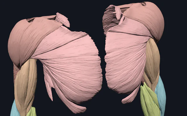
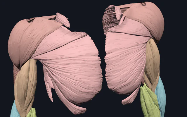
| Pectoralis major | Medial half of the clavicle; front of the manubrium; front of the body of the sternum; costal cartilages of the upper ribs; aponeurosis of the external oblique | Crest of the greater tubercle, the lateral lip of the intertubercular groove | Shoulder adduction, Medial rotation | Lateral and medial pectoral nerves (C5, C6, C7, C8, T1) |
| Pectoralis minor | Third rib near its cartilage; fourth rib near its cartilage; fifth rib near its cartilage | Coracoid process | Protraction | Medial pectoral nerve (C8, T1) |
| Serratus anterior | First rib; second rib; third rib; fourth rib; fifth rib; sixth rib; seventh rib; eighth rib | Front of the scapula’s medial border | Protraction, Upward rotation | Long thoracic nerve (C5, C6, C7) |
| Subclavius | First rib and its cartilage, where they meet | Groove under the middle third of the clavicle | The collarbone is drawn down and in | Nerve to subclavius (C5, C6) |
| Trapezius | External occipital protuberance and the superior nuchal line; ligamentum nuchae, to the cervical spines; spine of the seventh cervical vertebra; spine of the first thoracic vertebra; spine of the second thoracic vertebra; spine of the third thoracic vertebra; spines of the lower thoracic vertebrae, to the twelfth | Back of the lateral third of the clavicle; acromion and the upper lip of the spine of the scapula; tubercle at the medial end of the spine of the scapula | Elevation, Retraction, Upward rotation | Accessory nerve, cranial nerve XI, with C3 and C4 for sensation |
| Levator scapulae | Transverse process of the atlas; transverse process of the axis; transverse process of the third cervical vertebra; transverse process of the fourth cervical vertebra | Medial border of the scapula, from the superior angle to the spine | Elevation | Dorsal scapular nerve (C5), with branches of C3 and C4 |
| Rhomboid minor | Lower ligamentum nuchae; spine of the seventh cervical vertebra; spine of the first thoracic vertebra | Medial border of the scapula at the root of the spine | Retraction | Dorsal scapular nerve (C4, C5) |
| Rhomboid major | Spine of the second thoracic vertebra; spine of the third thoracic vertebra; spine of the fourth thoracic vertebra; spine of the fifth thoracic vertebra | Medial border of the scapula, from the spine to the inferior angle | Retraction | Dorsal scapular nerve (C4, C5) |
| Arm flexors | ||||
| Biceps brachii | Supraglenoid tubercle, long head; tip of the coracoid process, short head | Radial tuberosity; bicipital aponeurosis, into the forearm’s deep fascia | Elbow flexion, Supination | Musculocutaneous nerve (C5, C6) |
| Coracobrachialis | Tip of the coracoid process | Middle of the medial border of the humerus | Shoulder flexion | Musculocutaneous nerve (C6, C7) |
| Brachialis | Lower half of the front of the humerus | Coronoid process and the tuberosity of the ulna | Elbow flexion | Musculocutaneous nerve (C5, C6) |
| Brachioradialis | Upper two-thirds of the lateral supracondylar ridge of the humerus | Side of the base of the radius’s styloid process | Elbow flexion | Radial nerve (C5, C6) |
| Arm extensors | ||||
| Latissimus dorsi | Spines of the lower six thoracic vertebrae; thoracolumbar fascia, to the lumbar and sacral spines; back of the iliac crest; lower ribs | Floor of the intertubercular groove | Shoulder extension, Shoulder adduction | Thoracodorsal nerve (C6, C7, C8) |
| Triceps brachii | Infraglenoid tubercle, long head; back of the humerus above the radial groove, lateral head; back of the humerus below the radial groove, medial head | Olecranon | Elbow extension | Radial nerve (C6, C7, C8) |
| Anconeus | Back of the lateral epicondyle of the humerus | Side of the olecranon and the top of the back of the ulna | Elbow extension | Radial nerve (C7, C8) |
| Forearm rotators | ||||
| Supinator | Lateral epicondyle of the humerus; radial collateral and annular ligaments; supinator crest of the ulna | Side of the upper third of the radius | Supination | Deep branch of the radial nerve (C5, C6) |
| Pronator teres | Medial epicondyle of the humerus, humeral head; coronoid process of the ulna, ulnar head | Middle of the side of the radius | Pronation | Median nerve (C6, C7) |
| Pronator quadratus | Lower quarter of the front of the ulna | Lower quarter of the front of the radius | Pronation | Anterior interosseous nerve (C8, T1) |
| Muscles of mastication | ||||
| Masseter | Lower border of the zygomatic arch, zygomatic bone; lower border of the zygomatic arch, temporal bone | Angle and the outer surface of the ramus of the mandible | Jaw elevation | Masseteric nerve (V3) |
| Temporalis | Temporal fossa, temporal bone; temporal fossa, parietal bone; temporal fossa, frontal bone; temporal fossa, greater wing of the sphenoid; deep surface of the temporal fascia | Coronoid process of the mandible | Jaw elevation | Deep temporal nerves (V3) |
| Medial pterygoid | Lateral pterygoid plate of the sphenoid, its inner surface; pyramidal process of the palatine bone; tuberosity of the maxilla | Inner surface of the ramus and angle of the mandible | Jaw elevation | Nerve to medial pterygoid (V3) |
| Lateral pterygoid | Infratemporal surface of the greater wing of the sphenoid, upper head; outer surface of the lateral pterygoid plate, lower head | Pterygoid fovea on the neck of the mandible’s condyle; jaw joint’s disc and capsule | Protrusion | Nerve to lateral pterygoid (V3) |
| Neck | ||||
| Sternocleidomastoid | Front of the manubrium, sternal head; medial third of the clavicle, clavicular head | Mastoid process; lateral half of the superior nuchal line | Turning the head to the opposite side | Accessory nerve, cranial nerve XI, with C2 and C3 for sensation |
What the model leaves out
- Fascia, ligaments and aponeuroses. They are not segmented, so attachments to them are named in the card but not drawn.
- Some tendon ends. A few tendons stop short of the bone in the scan, which is what the threshold ladder and the pins are for.
- Every bone of a run. Where a muscle arises from a run of ribs, cartilages or vertebrae, as latissimus dorsi does from the lower six thoracic spines, its footprint is measured on one of them, which the card names, and By bone lists the muscle on the rest.
- One body. The meshes are one adult man’s. The landmarks are much the same in everyone, though how many ribs or vertebrae a muscle reaches varies from person to person, and the sizes and angles here are his.
- The hip and leg, and the muscles of the wrist and hand. The explorer covers the shoulder and arm, the jaw and the sternocleidomastoid; the regional muscle explorers have the rest of the muscles in 3D.
Common mistakes
- Reading contact as attachment. A muscle lies on the bones it crosses: the deltoid on the greater tubercle, supraspinatus on the clavicle. Only where it ends on the bone is an attachment.
- Quoting actions outside the anatomical position. Actions are stated with the body standing, arms by the sides and palms forward. Away from it a muscle can change sides: the clavicular part of pectoralis major flexes the arm, while the sternocostal part extends it back down from flexion.
More bones and muscles
The bones themselves are in the upper limb skeleton, lower limb skeleton and full skeleton explorers, and every muscle mesh of the region in the upper limb, back and head and neck muscle explorers. How a muscle makes force is the muscle twitch and tetanus simulator’s subject, and which muscles a lift uses is the muscles worked explorer’s, with a page for each lift in muscles worked by exercise. The skin and muscles each spinal root supplies are set out root by root in dermatomes by nerve root.
Common questions
What is the difference between a muscle’s origin and its insertion?
The origin is the attachment that usually stays fixed, often the end nearer the trunk, and the insertion the one that moves. Contraction pulls the insertion towards the origin, and the roles swap when the far end is the fixed one, as for latissimus dorsi in a pull-up, which pulls the trunk up to the arm.
What are the origins and insertions of the rotator cuff muscles?
Supraspinatus arises from the supraspinous fossa, infraspinatus from the infraspinous fossa and teres minor from the upper part of the scapula’s lateral border, and the three insert in that order down the facets of the greater tubercle of the humerus. Subscapularis runs from the subscapular fossa on the front of the scapula to the lesser tubercle (Gray, 1918).
Where does the deltoid originate and insert?
From the lateral third of the clavicle, the acromion and the spine of the scapula, to the deltoid tuberosity on the outer side of the humerus. Measured on this body, its origins cover 2.2 cm² of the clavicle, 2.4 cm² of the acromion and 6.7 cm² of the spine of the scapula, and its insertion 7.1 cm² of the humerus, centred 37 percent of the way down.
Which muscles attach to the humerus?
Of the muscles in this explorer, nine insert on the humerus: supraspinatus, infraspinatus, teres minor, subscapularis, teres major, latissimus dorsi, pectoralis major, coracobrachialis and the deltoid. Six arise from it: brachialis, two heads of triceps, anconeus, brachioradialis, supinator and the humeral head of pronator teres. Choose By bone and click the humerus to see them all painted.
What are the origins and insertions of the muscles of mastication?
Masseter runs from the zygomatic arch to the outer surface of the ramus and angle of the mandible, temporalis from the temporal fossa to the coronoid process, medial pterygoid from the lateral pterygoid plate, the palatine bone and the maxilla to the inner surface of the ramus and angle, and lateral pterygoid from the sphenoid to the neck of the condyle and the jaw joint’s disc.
Why are some attachments shown as a pin instead of a painted patch?
A patch is only painted where at least 25 mm² of bone lies within 3 mm of the muscle, in the right place on the bone. Some muscles stop up to about a centimetre short of the bone in the scan, as at several vertebral spines, so those sites are a hollow pin. Attachments to the ligamentum nuchae, the thoracolumbar fascia, the bicipital aponeurosis and the jaw joint’s disc are named but not drawn, because BodyParts3D does not segment those structures.
Is the 3D model male or female, and do attachments differ?
It is one adult male body. The attachment landmarks are the same in both sexes, while sizes and angles differ from person to person, so the areas measured here are this body’s and not a rule.