Ankle and Foot Skeleton Explorer
Turn all 26 bones of the ankle and foot in 3D. Click any bone to name it, isolate it, or slice through the model in standard anatomical views.
Visualiser
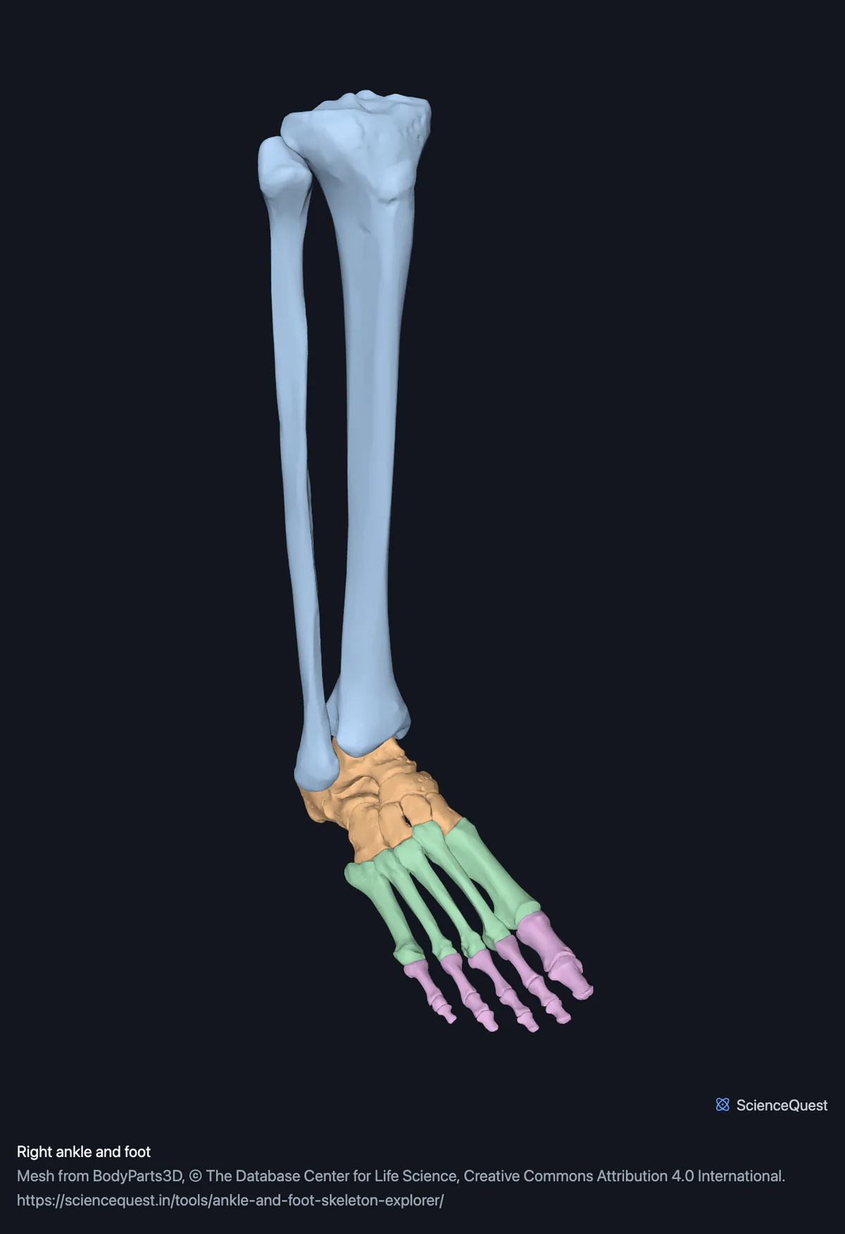
Right ankle and foot, 26 bones in 28 meshes
Checking this device for a stored copy.
Start here
- Search cuneiform, then switch on Isolate selection The cuboid and the lateral cuneiform look alike in a textbook. Turned on their own they are plainly different objects.
- Switch on Cut plane and drag it through the hindfoot Separates the ankle joint proper from the subtalar joint underneath it, which no single view shows.
- Select the talus and look at how much of it is joint surface About three-fifths of it, which is the reason a displaced fracture there is serious.
Bone only. No ligaments, tendons, muscle, cartilage, vessels or nerves, for two different reasons. The only ligaments BodyParts3D publishes are the inguinal and long plantar ligaments of each side, so the ones that hold an ankle together are simply not available, and the long plantar ligaments are in the full skeleton model rather than this one. The foot’s muscles are published, and they are in the lower limb muscle model rather than this one. Either way this teaches you the skeleton and not an ankle sprain.
- 26 bones
- 68k triangles
- 360 KB, on request
- 0.00313 mm worst error per axis
- 2 leg
- 7 tarsals
- 5 metatarsals
- 14 phalanges
The right tibia and right fibula load with them for context and are not counted above. 28 meshes load in total, and every one of them is listed, nameable and selectable.
A foot has 26 bones: seven tarsals, five metatarsals and fourteen phalanges. The tibia and fibula are loaded with them because an ankle joint cannot be read without the leg it hangs from, so 28 meshes arrive and 26 of them are bones of the foot.
Where this comes from
Segmented from imaging of one adult male body, not drawn by an artist, so the shapes are measurements. Two consequences worth knowing: it is one person’s anatomy rather than an average, and faint stepping on some surfaces is the resolution of the original scan rather than a rendering fault.
Meshes from BodyParts3D, © The Database Center for Life Science, under Creative Commons Attribution 4.0 International. Names and identifiers are the dataset’s own: Foundational Model of Anatomy identifiers and English labels, as its parts list gives them. Converted meshes are shared under the same licence.
Changes made: Meshes were converted from binary STL to an indexed mesh with 16-bit quantised vertex positions, and surface normals were recomputed from the geometry. No vertex was moved other than by that quantisation, whose worst-case error on any one coordinate is reported on the page.
Latin terms for 8 of the 28 structures come from English Wikipedia anatomy infoboxes, under Creative Commons Attribution-ShareAlike 4.0, joined by Wikidata property P1402, Foundational Model of Anatomy identifier. Only a term a source states for that structure is used, never one constructed from a family name, which is why the rest carry no Latin rather than a guess.
All 28 structures
Every structure the model contains, group by group. Of these, 26 are bones of this region. Each name opens the viewer with that structure selected, so a link can point at one structure rather than at the whole region.
Leg 2
Tarsals 7
- right talus talus
- right calcaneus calcaneus, calcaneum, os calcis
- navicular bone of right foot os naviculare
- right cuboid bone os cuboideum
- right medial cuneiform bone
- right intermediate cuneiform bone
- right lateral cuneiform bone
Metatarsals 5
- right first metatarsal bone os metatarsale I
- right second metatarsal bone os metatarsale II
- right third metatarsal bone
- right fourth metatarsal bone os metatarsale IV
- right fifth metatarsal bone os metatarsale V
Phalanges 14
- proximal phalanx of right big toe
- distal phalanx of right big toe
- proximal phalanx of right second toe
- middle phalanx of right second toe
- distal phalanx of right second toe
- proximal phalanx of right third toe
- middle phalanx of right third toe
- distal phalanx of right third toe
- proximal phalanx of right fourth toe
- middle phalanx of right fourth toe
- distal phalanx of right fourth toe
- proximal phalanx of right little toe
- middle phalanx of right little toe
- distal phalanx of right little toe
Citing this tool
Last updated . Add the date you accessed it as well, which a citation of a page that can change asks for. If a specific view matters, cite the page address as it stands in your browser instead: it records what is selected and shown.
The seven tarsals, in order
Anyone can name the talus and the calcaneus. The other five defeat people because a flat diagram teaches their position on the page rather than their shape.
Medial to lateral, front to back:
- Medial, intermediate and lateral cuneiform, in a row in front of the navicular.
- Cuboid, lateral to those, in front of the calcaneus.
- Navicular and talus behind them, then the calcaneus.
That row is also why the second metatarsal is recessed: it is mortised between the medial and lateral cuneiforms, which makes it the most rigid of the five and is why a Lisfranc injury is described around it.
The ankle is two joints
- Talocrural
- The talus held in a mortise between tibia and fibula. Points the foot up and down.
- Subtalar
- The talus against the calcaneus, below it. Turns the sole inward and outward.
The talus is unusual in having no muscle attached to it at all. It is held by its shape and by ligaments, and so much of its surface is articular that there is little room for vessels to enter.
Common questions
How many bones are in the foot and ankle, and why do counts differ?
A foot has 26 bones: seven tarsals, five metatarsals and fourteen phalanges. That is the figure this page leads with, and it is the one you will find in Gray’s or in the NIH’s own anatomy reference. This model loads 28 meshes, because the tibia and fibula come with them: the ankle joint is the talus meeting those two bones, and you cannot show that joint without them. They are listed and selectable like everything else, they are simply not counted as bones of the foot. Higher counts elsewhere usually add the sesamoid bones under the big toe, which nearly everyone has. BodyParts3D does publish those, as one combined mesh per foot rather than as two separate bones, so this region leaves them out. Any source quoting a single figure without saying what it includes is being imprecise, which is why both numbers are on this page.
Why does the big toe have only two bones when every other toe has three?
Because that is how the hallux is built, and it is not an omission in the model. Every other toe has a proximal, a middle and a distal phalanx. The big toe has a proximal and a distal one only, which is part of why it is so much stronger: fewer joints in the same length means less to stabilise when you push off. The same pattern holds in the hand, where the thumb has two phalanges and the other fingers have three.
Are these bones a real person, or a model someone drew?
They are segmented from imaging of one adult male body, published as BodyParts3D by the Database Center for Life Science in Japan. That means the shapes are real measurements rather than an artist’s interpretation, which is the reason to prefer them, but it also means they are one individual’s bones and not an average. Real feet vary a good deal, particularly in the height of the arch and the relative lengths of the metatarsals. The surfaces also show faint stepping in places, which is the resolution of the original scan showing through rather than a rendering fault.
Why are the bones different colours when real bone is all one colour?
Because seven similarly shaped ivory bones packed together are almost impossible to tell apart on a screen. The colour-by-group option gives each anatomical group its own hue, which is the convention printed atlases have used for a century for exactly this reason. It is a teaching device and not a claim about appearance, so there is also a bone option that paints everything the off-white of a dry specimen. The viewer says which one is showing. The underlying data carries no colour at all: the file format stores only geometry.
Why does the model download only when I ask for it?
Because it is about 360 kilobytes of mesh plus the 3D engine, and most visitors to a page like this are reading rather than rotating. Loading it automatically would slow the page for everyone to benefit the few who want it, and page speed feeds search ranking, so it would also cost the page its readers. Pressing the button fetches it with a progress bar, and the checksum is verified before anything is drawn so a half-finished download cannot show you an incomplete skeleton.