Medial cuneiform bone
The medial cuneiform is the largest of the three wedge-shaped cuneiform bones of the midfoot. It lies on the medial side of the foot, between the navicular and the base of the first metatarsal. (Gray’s Anatomy (1918), First cuneiform bone, p. 270; StatPearls, Foot and Ankle)
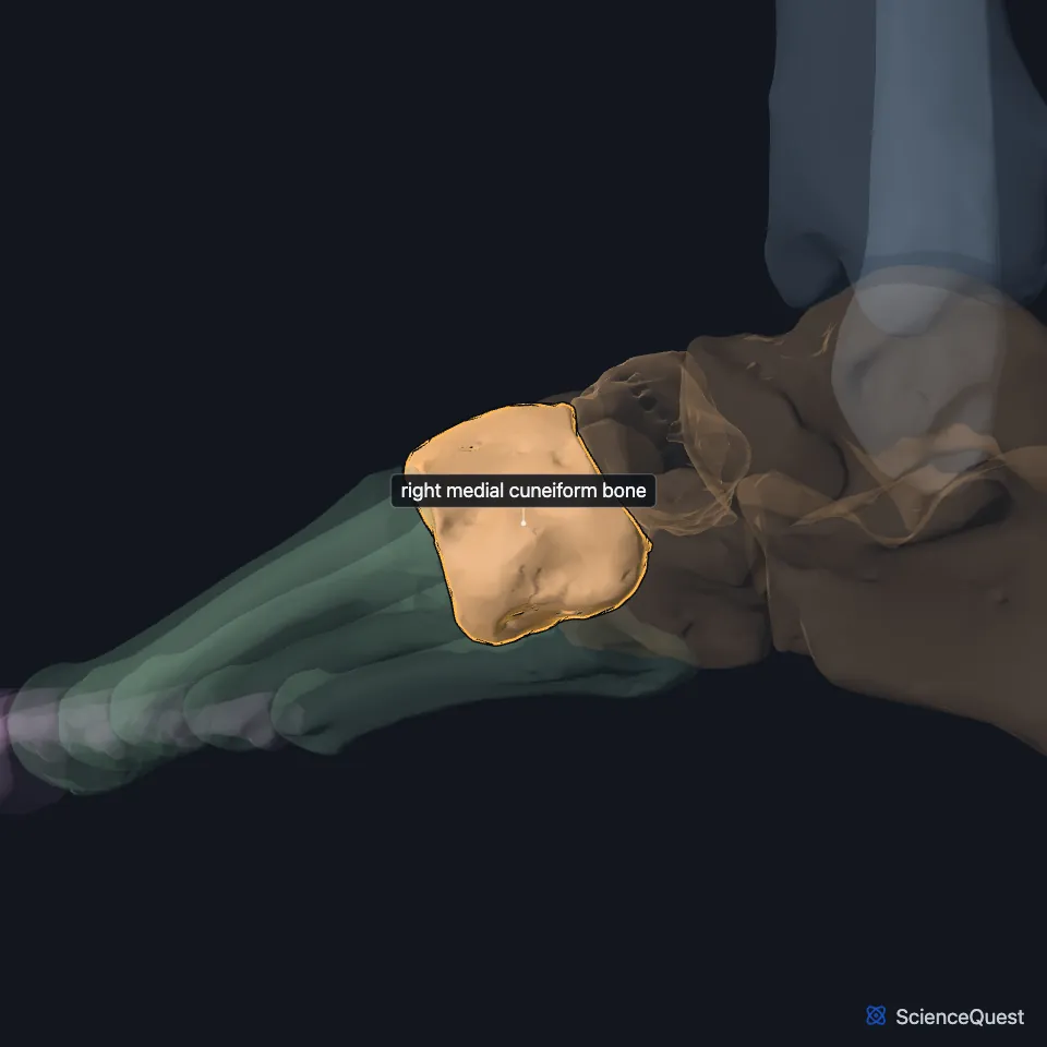
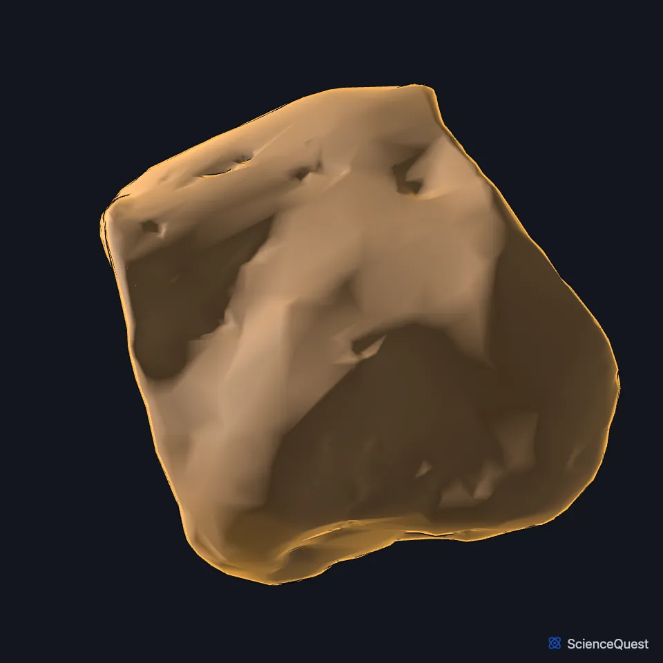
Open it in the Ankle and Foot Skeleton Explorer See it alone in 3D
Where is the medial cuneiform?
It sits in front of the navicular and behind the first metatarsal, and its medial surface lies under the skin. Its wedge stands with the broad base down, on the sole, and the narrow edge up. (Gray’s Anatomy (1918), First cuneiform bone, p. 270)
Which bones does the medial cuneiform meet?
Four: the navicular, the intermediate cuneiform, and the first and second metatarsals. With the first metatarsal it makes the medial column of the tarsometatarsal joints. (Gray’s Anatomy (1918), First cuneiform bone, p. 270; StatPearls, Metatarsal Bones)
The Lisfranc ligament runs from its lateral surface to the medial base of the second metatarsal. It is the main stabilizer of the midfoot, holding the medial and middle columns together. (StatPearls, Lisfranc Dislocation)
Which tendons insert on the medial cuneiform?
Tibialis anterior inserts on its medial and plantar surfaces and on the base of the first metatarsal. Because the muscle arises on the lateral tibia, its pull lifts the medial arch, the medial cuneiform with the first metatarsal, navicular and talus, into inversion. (StatPearls, Medial Longitudinal Arch of the Foot and Tibialis Anterior Muscles)
Fibularis longus crosses the sole from the lateral side, under the cuboid, to insert on the medial cuneiform and the base of the first metatarsal. A slip of tibialis posterior reaches the tuberosity on its sole surface. (StatPearls, Arches of the Foot and Calf Peroneus Longus Muscle; Gray’s Anatomy (1918), First cuneiform bone, p. 270)
What injuries and variants involve the medial cuneiform?
A Lisfranc injury is a disruption of the joint between the medial cuneiform and the base of the second metatarsal. Pain on pressing between those two bones should raise the suspicion of a torn Lisfranc ligament. (StatPearls, Metatarsal Bones and Lisfranc Dislocation)
Some people have a medial cuneiform in two pieces, a bipartite bone that is usually found by chance and causes no symptoms. (StatPearls, Foot and Ankle)
| Feature | Detail and source |
|---|---|
| Articulates with | Navicular, intermediate cuneiform, first and second metatarsals (Gray’s Anatomy (1918), First cuneiform bone, p. 270) |
| Tendons inserting | Tibialis anterior, fibularis longus, part of tibialis posterior (StatPearls, Medial Longitudinal Arch of the Foot and Arches of the Foot; Gray’s Anatomy (1918), First cuneiform bone, p. 270) |
| Older name | First, or internal, cuneiform (Gray’s Anatomy, 1918) (Gray’s Anatomy (1918), First cuneiform bone, p. 270) |
Touches in this model
Measured on the BodyParts3D meshes: surfaces within 2 mm of the medial cuneiform bone, nearest first, 12 of 13.
- Right first metatarsal bone in contact
- Right intermediate cuneiform bone in contact
- Right second metatarsal bone in contact
- Lateral head of right flexor hallucis brevis in contact
- Right extensor hallucis brevis in contact
- Right fibularis longus in contact
- Right long plantar ligament in contact
- Right tibialis posterior in contact
- Skin in contact
- Navicular bone of right foot 0.2 mm away
- Right extensor hallucis longus 1.3 mm away
- Medial head of right flexor hallucis brevis 1.6 mm away
Sources
Sources read
- Gray’s Anatomy of the Human Body, 20th US edition (1918), edited by Warren H. Lewis, Osteology, The Tarsus, The First Cuneiform Bone (pp. 270 to 271); 1918 names: first cuneiform for medial cuneiform, second cuneiform for intermediate cuneiform. Henry Gray; Warren H. Lewis (editor). Public domain. Read .
- StatPearls, Anatomy, Bony Pelvis and Lower Limb: Foot and Ankle. Jennifer Ficke; Adegbenro O. Fakoya; Doug W. Byerly. Last update . CC BY-NC-ND 4.0. Read .
- StatPearls, Anatomy, Bony Pelvis and Lower Limb: Metatarsal Bones. Bradley Lezak; James La Rose; Dustin H. Massel. Last update . CC BY-NC-ND 4.0. Read .
- StatPearls, Lisfranc Dislocation. Jonathan M. Stern; Rachel Bergman; Swapnil Singh. Last update . CC BY-NC-ND 4.0. Read .
- StatPearls, Anatomy, Bony Pelvis and Lower Limb: Medial Longitudinal Arch of the Foot. Deepika Babu; Bruno Bordoni. Last update . CC BY-NC-ND 4.0. Read .
- StatPearls, Anatomy, Bony Pelvis and Lower Limb: Tibialis Anterior Muscles. Pallavi Juneja; John B. Hubbard. Last update . CC BY-NC-ND 4.0. Read .
- StatPearls, Anatomy, Bony Pelvis and Lower Limb: Arches of the Foot. Hiteshkumar M. Chauhan; Muhammad Taqi. Last update . CC BY-NC-ND 4.0. Read .
- StatPearls, Anatomy, Bony Pelvis and Lower Limb: Calf Peroneus Longus Muscle. Bradley Lezak; Matthew A. Varacallo. Last update . CC BY-NC-ND 4.0. Read .
The values are facts and free to use; this page’s selection and presentation are © 2026 ScienceQuest.