Tibialis posterior
The tibialis posterior is the deepest muscle of the deep posterior compartment of the leg. It plantarflexes the ankle, inverts the foot and is the main stabilizer of the medial longitudinal arch. (StatPearls, Tibialis Posterior Muscle and Leg Posterior Compartment)
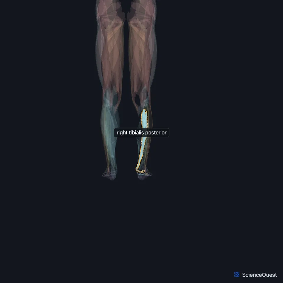
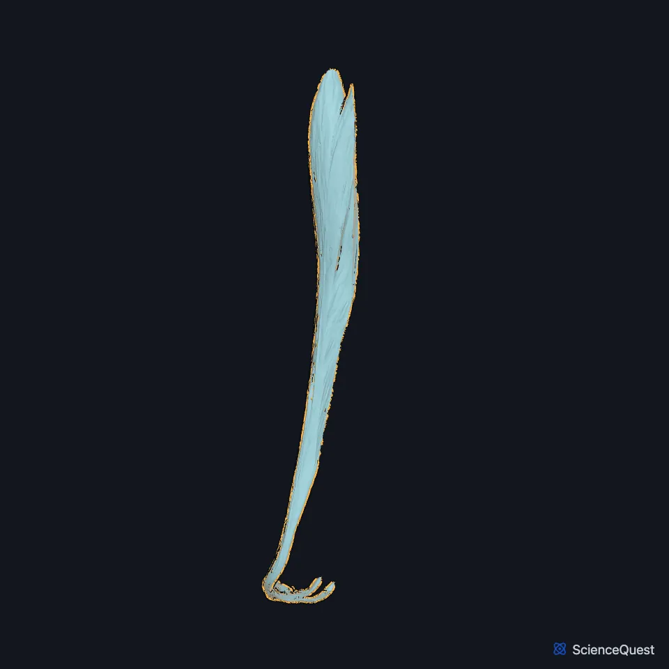
Open it in the Lower Limb Muscles Explorer See it alone in 3D
Where is the tibialis posterior?
It lies between the flexor digitorum longus and the flexor hallucis longus, deeper than both. It arises from the back of the interosseous membrane, the upper back of the tibia and the middle of the back of the fibula. (StatPearls, Tibialis Posterior Muscle; Gray’s Anatomy (1918), Posterior Crural Muscles, deep group)
Its upper end splits into two pointed processes. The anterior tibial artery passes forward between them and through the upper interosseous membrane to the front of the leg. (Gray’s Anatomy (1918), Posterior Crural Muscles, deep group; StatPearls, Tibial Nerve)
Where does its tendon go?
Its tendon passes behind the medial malleolus with the tendons of the flexor digitorum longus and flexor hallucis longus, the posterior tibial artery and the tibial nerve. It runs under the flexor retinaculum in a synovial-lined tunnel. (StatPearls, Tibialis Posterior Muscle)
In the sole it splits into three parts. The main part inserts on the tuberosity of the navicular and the medial cuneiform, the plantar part on the second to fourth metatarsal bases, the other two cuneiforms and the cuboid, and a small recurrent part on the sustentaculum tali of the calcaneus. (StatPearls, Tibialis Posterior Muscle)
Gray’s 1918 notes a sesamoid fibrocartilage in the tendon where it runs under the plantar calcaneonavicular ligament. (Gray’s Anatomy (1918), Posterior Crural Muscles, deep group)
What does the tibialis posterior do?
With the tibialis anterior it is a primary inverter of the foot, at the midtarsal and subtalar joints. It also plantarflexes the foot, a secondary role beside the calf muscles. (StatPearls, Tibialis Posterior Muscle)
In walking it holds up the arch, controls midfoot pronation in midstance and locks the transverse tarsal joint at push-off. That lock turns the midfoot into a rigid lever for the calf muscles. (StatPearls, Leg Posterior Compartment)
Failure of its tendon lets the medial arch fall. Posterior tibial tendon insufficiency is the most common cause of flatfoot acquired in adulthood. (StatPearls, Tibialis Posterior Muscle)
Its main antagonists are the anterior compartment muscles, which dorsiflex the foot. (StatPearls, Tibialis Posterior Muscle)
What supplies the tibialis posterior?
The tibial nerve and the posterior tibial artery run down its back surface, deep to the soleus, and both supply it. (StatPearls, Tibialis Posterior Muscle)
The tibial nerve follows its tendon through the tarsal tunnel behind the medial malleolus, and compression of the nerve there causes tarsal tunnel syndrome. (StatPearls, Tibialis Posterior Muscle)
Does the tibialis posterior vary?
Its tendon may form three separate tendons, main, plantar and recurrent, or one wide tendon that reaches the same bones. (StatPearls, Tibialis Posterior Muscle)
Where the sources differ
Sources give different roots for its nerve: L4 to L5 in the StatPearls popliteus article, the fifth lumbar and first sacral nerves in Gray’s 1918. (StatPearls, Popliteus Muscle; Gray’s Anatomy (1918), Posterior Crural Muscles, deep group)
| Feature | Detail and source |
|---|---|
| Origin | Back of the interosseous membrane; upper back of the tibia; middle of the back of the fibula (StatPearls, Tibialis Posterior Muscle) |
| Insertion | Navicular tuberosity and medial cuneiform, with slips to the other tarsals, metatarsal bases 2 to 4 and the sustentaculum tali (StatPearls, Tibialis Posterior Muscle) |
| Action | Plantarflexes and inverts the foot; supports the medial arch (StatPearls, Tibialis Posterior Muscle) |
| Nerve | Tibial nerve (StatPearls, Tibialis Posterior Muscle) |
Touches in this model
Measured on the BodyParts3D meshes: surfaces within 2 mm of the tibialis posterior, nearest first, 12 of 22.
- Lateral head of right flexor hallucis brevis in contact
- Medial head of right flexor hallucis brevis in contact
- Oblique head of right adductor hallucis in contact
- Right abductor hallucis in contact
- Right extensor digitorum longus in contact
- Right fibularis longus in contact
- Right flexor digitorum longus in contact
- Right flexor hallucis longus in contact
- Right soleus in contact
- Interosseous membrane of right leg in contact
- Navicular bone of right foot in contact
- Right first metatarsal bone in contact
Sources
Sources read
- StatPearls, Anatomy, Bony Pelvis and Lower Limb: Tibialis Posterior Muscle. Nicholas M. Corcoran; Matthew A. Varacallo. Last update . CC BY-NC-ND 4.0. Read .
- StatPearls, Anatomy, Bony Pelvis and Lower Limb: Leg Posterior Compartment. Rami Abuqubo; Steven B. Graefe; James La Rose. Last update . CC BY-NC-ND 4.0. Read .
- Anatomy of the Human Body (Gray’s Anatomy), 20th US edition, 1918, Myology, The Muscles and Fasciae of the Leg, 2. The Posterior Crural Muscles, the deep group, pages 483 to 486; Internet Archive scan of the 1918 Lea and Febiger printing, terminology of 1918. Henry Gray; edited by Warren H. Lewis. Public domain. Read .
- StatPearls, Anatomy, Bony Pelvis and Lower Limb: Tibial Nerve. Sohil S. Desai; James La Rose. Last update . CC BY-NC-ND 4.0. Read .
- StatPearls, Anatomy, Bony Pelvis and Lower Limb: Popliteus Muscle. Scott Hyland; Matthew A. Varacallo. Last update . CC BY-NC-ND 4.0. Read .
The values are facts and free to use; this page’s selection and presentation are © 2026 ScienceQuest.