Tibialis anterior
The tibialis anterior is the largest of the four muscles in the anterior compartment of the leg. It dorsiflexes and inverts the foot. (StatPearls, Tibialis Anterior Muscles)
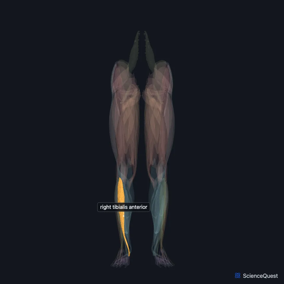
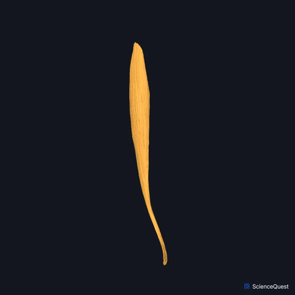
Open it in the Lower Limb Muscles Explorer See it alone in 3D
Where is the tibialis anterior?
It runs down the lateral side of the tibia, fleshy and thick in its upper part and tendinous lower down. Extensor digitorum longus runs along its lateral side, and extensor hallucis longus sits between the two. (OpenStax A&P 2e, 11.6; Gray’s Anatomy (1918), Anterior Crural Muscles)
It is the most medial muscle of the compartment, and you can see its tendon on the front of the ankle. (StatPearls, Leg Anterior Compartment)
Where does the tibialis anterior attach?
It arises from the lateral condyle and upper two-thirds of the lateral surface of the tibia, the interosseous membrane, the deep surface of the crural fascia and the septum it shares with extensor digitorum longus. (StatPearls, Tibialis Anterior Muscles)
Its tendon begins in the lower third of the leg, crosses the front of the ankle and the top of the foot, and inserts on the medial cuneiform and the base of the first metatarsal. It is the most medial tendon of the ankle and foot. (StatPearls, Tibialis Anterior Muscles)
In most people the tendon passes under the extensor retinaculum, which holds it in place. (StatPearls, Tibialis Anterior Muscles)
What does the tibialis anterior do?
It is the strongest dorsiflexor of the foot, the movement that brings the top of the foot toward the leg. Dorsiflexion lifts the foot clear of the ground in the swing phase of walking. (StatPearls, Tibialis Anterior Muscles; OpenStax A&P 2e, 9.5)
With tibialis posterior it is a main inverter of the foot. Its pull into inversion is strong enough that the lateral compartment muscles must evert the foot for it to dorsiflex without inversion. (StatPearls, Tibialis Anterior Muscles)
Through its insertion on the inner side of the foot it supports the medial longitudinal arch, which the calcaneus, talus, navicular, the three cuneiforms and the first three metatarsals form. (StatPearls, Tibialis Anterior Muscles)
What supplies the tibialis anterior?
The deep fibular nerve, also called the deep peroneal nerve, supplies it and the other three muscles of the compartment. The nerve is a branch of the common fibular nerve. (StatPearls, Tibialis Anterior Muscles and Calf Deep Peroneal Nerve)
The nerve and the anterior tibial artery run between it and extensor digitorum longus higher up, and between it and extensor hallucis longus lower down. (StatPearls, Tibialis Anterior Muscles)
The anterior tibial artery supplies the muscle, and the medial tarsal branches of the dorsalis pedis artery supply its tendon. (StatPearls, Tibialis Anterior Muscles)
Its antagonists are the muscles of the posterior compartment, among them the gastrocnemius and soleus, which plantarflex the foot. (StatPearls, Tibialis Anterior Muscles and Leg Posterior Compartment)
Does the tibialis anterior vary?
In a quarter of cases the superficial and deep layers of the superior extensor retinaculum form a separate tunnel for its tendon. (StatPearls, Tibialis Anterior Muscles)
Where the sources differ
Sources differ on the spinal nerves in the common fibular nerve, the parent of its nerve: L4 to S1 in the StatPearls tibialis anterior article, L4 to S2 in its deep fibular nerve article. (StatPearls, Tibialis Anterior Muscles and Calf Deep Peroneal Nerve)
| Feature | Detail and source |
|---|---|
| Origin | Lateral condyle and upper two-thirds of the lateral tibia; interosseous membrane; crural fascia (StatPearls, Tibialis Anterior Muscles) |
| Insertion | Medial cuneiform and base of the first metatarsal (StatPearls, Tibialis Anterior Muscles) |
| Action | Dorsiflexes and inverts the foot (StatPearls, Tibialis Anterior Muscles) |
| Nerve | Deep fibular (deep peroneal) nerve (StatPearls, Tibialis Anterior Muscles) |
Touches in this model
Measured on the BodyParts3D meshes: surfaces within 2 mm of the tibialis anterior, nearest first.
- Medial head of right flexor hallucis brevis in contact
- Right extensor digitorum longus in contact
- Right extensor hallucis longus in contact
- Right iliotibial tract in contact
- Interosseous membrane of right leg in contact
- Right tibia in contact
- Skin in contact
- Navicular bone of right foot 1.0 mm away
- Right first metatarsal bone 2.0 mm away
Sources
- StatPearls, Anatomy, Bony Pelvis and Lower Limb: Tibialis Anterior Muscles. Pallavi Juneja; John B. Hubbard. Last update August 28, 2023. CC BY-NC-ND 4.0. Read 2026-10-25.
- OpenStax Anatomy and Physiology 2e, section 11.6, Appendicular Muscles of the Pelvic Girdle and Lower Limbs. CC BY-NC-SA 4.0. Read 2026-10-25.
- Anatomy of the Human Body (Gray’s Anatomy), 20th US edition, 1918, Myology, The Muscles and Fasciae of the Leg, 1. The Anterior Crural Muscles, pages 480 to 482; Internet Archive scan of the 1918 Lea and Febiger printing, terminology of 1918. Henry Gray; edited by Warren H. Lewis. Public domain. Read 2026-10-25.
- StatPearls, Anatomy, Bony Pelvis and Lower Limb: Leg Anterior Compartment. Bradley Lezak; Marjorie V. Launico. Last update June 8, 2026. CC BY-NC-ND 4.0. Read 2026-10-25.
- OpenStax Anatomy and Physiology 2e, section 9.5, Types of Body Movements. CC BY-NC-SA 4.0. Read 2026-10-25.
- StatPearls, Anatomy, Bony Pelvis and Lower Limb: Calf Deep Peroneal Nerve (Deep Fibular Nerve). Lane L. Palmisano; Marjorie V. Launico. Last update August 9, 2025. CC BY-NC-ND 4.0. Read 2026-10-25.
- StatPearls, Anatomy, Bony Pelvis and Lower Limb: Leg Posterior Compartment. Rami Abuqubo; Steven B. Graefe; James La Rose. Last update May 13, 2026. CC BY-NC-ND 4.0. Read 2026-10-25.
The values are facts and free to use; this page’s selection and presentation are © 2026 ScienceQuest.