Gastrocnemius
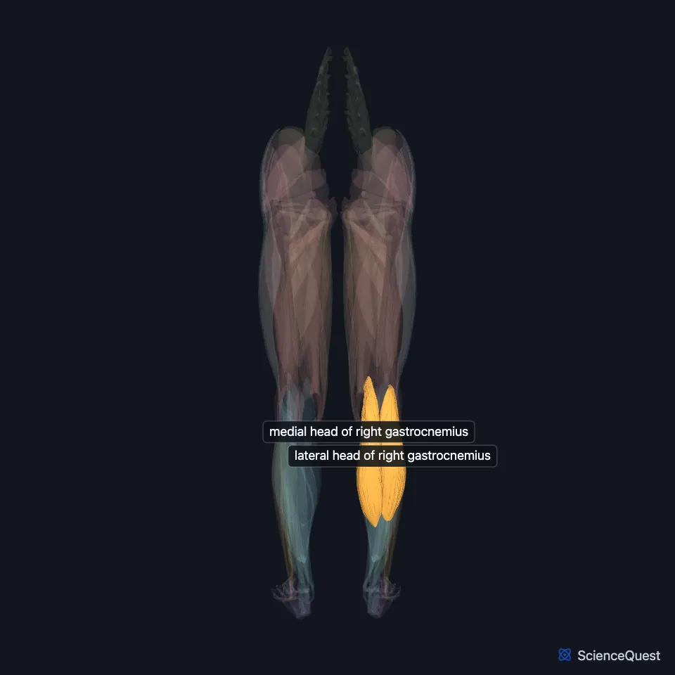
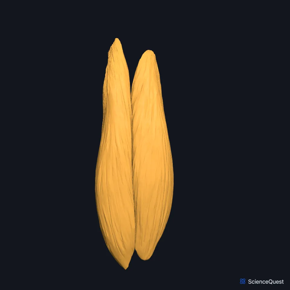
The gastrocnemius is the most superficial calf muscle and forms most of the calf. It has a medial and a lateral head, and with the soleus it forms the triceps surae. (OpenStax A&P 2e, 11.6; StatPearls, Gastrocnemius Muscle; Gray’s Anatomy (1918), Posterior Crural Muscles)
Open it in the Lower Limb Muscles Explorer See it alone in 3D
Where is the gastrocnemius?
Its two heads arise from the medial and lateral condyles of the femur, and it inserts on the calcaneus through the calcaneal tendon, so it crosses both the knee and the ankle. (StatPearls, Gastrocnemius Muscle)
The medial and lateral heads form the lower inner and lower outer borders of the popliteal fossa behind the knee. (StatPearls, Tibial Nerve)
The broad, flat soleus lies just in front of it. The small plantaris lies between the two, and its long thin tendon crosses obliquely between them. (OpenStax A&P 2e, 11.6; StatPearls, Calf; Gray’s Anatomy (1918), Posterior Crural Muscles)
What are the parts of the gastrocnemius?
The medial head is larger, thicker and wider than the lateral head. (StatPearls, Leg Posterior Compartment and Gastrocnemius Muscle)
In the middle third of the leg the heads unite in a broad aponeurosis, which continues down as the calcaneal tendon. It is the thickest and strongest tendon in the body and inserts on the middle of the back of the calcaneus. (StatPearls, Gastrocnemius Muscle; OpenStax A&P 2e, 11.6; Gray’s Anatomy (1918), Posterior Crural Muscles)
What does the gastrocnemius do?
It plantarflexes the ankle, lifting the heel, and helps flex the knee. (StatPearls, Gastrocnemius Muscle; OpenStax A&P 2e, 9.5; Gray’s Anatomy (1918), Posterior Crural Muscles)
It plantarflexes the ankle when the knee is straight; the soleus does so whatever the position of the knee. (StatPearls, Leg Posterior Compartment)
Most of its fibers are anaerobic, white fibers, and quick, forceful efforts recruit it by preference. Standing and walking rely mainly on the soleus. (StatPearls, Gastrocnemius Muscle)
What supplies the gastrocnemius?
The tibial nerve supplies it. The nerve descends into the popliteal fossa between its two heads, in front of the soleus. (StatPearls, Gastrocnemius Muscle)
The sural arteries, branches of the popliteal artery in the popliteal fossa, supply both heads and the soleus. (StatPearls, Gastrocnemius Muscle)
In walking it works with the quadriceps at first contact and at push-off, when the knee is near full extension: it resists forward slide of the tibia while the quadriceps extend the knee. (StatPearls, Gastrocnemius Muscle)
Does the gastrocnemius vary?
A small sesamoid bone, the fabella, sits in the origin of the lateral head in 10% to 30% of people. (StatPearls, Gastrocnemius Muscle)
Where the sources differ
Sources give different rates for a missing plantaris, the muscle between the gastrocnemius and the soleus. OpenStax finds none in about 7% of cadaver dissections, and the StatPearls calf article says it is absent in up to 10% of people. The StatPearls gastrocnemius article gives about 10% in one place and, in another, says the plantaris is present in only a small percentage of people. (OpenStax A&P 2e, 11.6; StatPearls, Calf and Gastrocnemius Muscle)
| Feature | Detail and source |
|---|---|
| Origin | Medial head: medial condyle of the femur. Lateral head: lateral condyle of the femur (StatPearls, Leg Posterior Compartment) |
| Insertion | Calcaneus, through the calcaneal tendon (StatPearls, Gastrocnemius Muscle) |
| Action | Plantarflexes the ankle; helps flex the knee (StatPearls, Gastrocnemius Muscle) |
| Nerve | Tibial nerve (StatPearls, Gastrocnemius Muscle) |
Touches in this model
Measured on the BodyParts3D meshes: surfaces within 2 mm of the gastrocnemius, nearest first, 12 of 15.
- Long head of right biceps femoris in contact
- Right adductor magnus in contact
- Right calcaneal tendon in contact
- Right plantaris in contact
- Right popliteus in contact
- Right semimembranosus in contact
- Right semitendinosus in contact
- Right soleus in contact
- Short head of right biceps femoris in contact
- Right femur in contact
- Right tibia in contact
- Skin in contact
Sources
- OpenStax Anatomy and Physiology 2e, section 11.6, Appendicular Muscles of the Pelvic Girdle and Lower Limbs. CC BY-NC-SA 4.0. Read 2026-10-25.
- StatPearls, Anatomy, Bony Pelvis and Lower Limb: Gastrocnemius Muscle. Bruno Bordoni; Matthew A. Varacallo. Last update April 11, 2026. CC BY-NC-ND 4.0. Read 2026-10-25.
- Anatomy of the Human Body (Gray’s Anatomy), 20th US edition, 1918, Myology, The Muscles and Fasciae of the Leg, 2. The Posterior Crural Muscles, superficial group, pages 482 to 485; Internet Archive scan of the 1918 Lea and Febiger printing, terminology of 1918. Henry Gray; edited by Warren H. Lewis. Public domain. Read 2026-10-25.
- StatPearls, Anatomy, Bony Pelvis and Lower Limb: Tibial Nerve. Sohil S. Desai; James La Rose. Last update September 14, 2026. CC BY-NC-ND 4.0. Read 2026-10-25.
- StatPearls, Anatomy, Bony Pelvis and Lower Limb: Calf. Justin T. Binstead; Matthew A. Varacallo. Last update January 31, 2026. CC BY-NC-ND 4.0. Read 2026-10-25.
- StatPearls, Anatomy, Bony Pelvis and Lower Limb: Leg Posterior Compartment. Rami Abuqubo; Steven B. Graefe; James La Rose. Last update May 13, 2026. CC BY-NC-ND 4.0. Read 2026-10-25.
- OpenStax Anatomy and Physiology 2e, section 9.5, Types of Body Movements. CC BY-NC-SA 4.0. Read 2026-10-25.
The values are facts and free to use; this page’s selection and presentation are © 2026 ScienceQuest.